Primers:
| 1642 | CARM1_FWD | TGGTTATCAACAGCCCCGAC | JH | 5/21/2015 | 20 | 55 | O.lurida | Histone-arginine methyltransferase CARM1 (EC 2.1.1.-) (EC 2.1.1.125) (Coactivator-associated arginine methyltransferase 1) (Protein arginine N-methyltransferase 4) | Q6DC04 | |
| 1641 | CARM1_REV | GTTGTTGACCCCAGGAGGAG | JH | 5/21/2015 | 20 | 55 | O.lurida | Histone-arginine methyltransferase CARM1 (EC 2.1.1.-) (EC 2.1.1.125) (Coactivator-associated arginine methyltransferase 1) (Protein arginine N-methyltransferase 4) | Q6DC04 |
Reagent Table:
| Volume | Reactions X58 | |
| Ssofast Evagreen MM | 10 | 580 |
| FWD Primer | 0.5 | 29 |
| REV Primer | 0.5 | 29 |
| Nuclease Free H2O | 8 | 464 |
| cDNA | 1 |
- Added reagents from greatest to least volume
- Vortexed
- Centrifuged briefly
- Pipetted 19 ul Master Mix to each tube
- Pipetted appropriate cDNA sample to each tube
- Centrifuged plate at 2000 rpm for 1 minute
- Ran Program Below
Program:
| Step | Temperature | Time |
| Initiation | 95 C | 10 min |
| Elongation | 95 C | 30 sec |
| 60 C | 1 min | |
| Read | ||
| 72 C | 30 sec | |
| Read | ||
| Repeat Elongation 39 times | ||
| Termination | 95 C | 1 min |
| 55 C | 1 sec | |
| Melt Curve Manual ramp 0.2C per sec Read 0.5 C | 55 - 95 C | 30 sec |
| 21 C | 10 min | |
| End |
Plate Layout:
| 1 | 2 | 3 | 4 | 5 | 6 | 7 |
| DNased 42215 HC1 | DNased 42215 NC1 | DNased 42215 SC1 | DNased 42215 HT1 1 | DNased 42215 NT1 1 | DNased 42215 ST1 1 | NTC |
| DNased 42215 HC2 | DNased 42215 NC2 | DNased 42215 SC2 | DNased 42215 HT1 2 | DNased 42215 NT1 2 | DNased 42215 ST1 2 | NTC |
| DNased 42215 HC3 | DNased 42215 NC3 | DNased 42215 SC3 | DNased 42215 HT1 3 | DNased 42215 NT1 3 | DNased 42215 ST1 3 | NTC |
| DNased 42215 HC4 | DNased 42215 NC4 | DNased 42215 SC4 | DNased 42215 HT1 4 | DNased 42215 NT1 4 | DNased 42215 ST1 4 | NTC |
| DNased 42215 HC5 | DNased 42215 NC5 | DNased 42215 SC5 | DNased 42215 HT1 5 | DNased 42215 NT1 5 | DNased 42215 ST1 5 | |
| DNased 42215 HC6 | DNased 42215 NC6 | DNased 42215 SC6 | DNased 42215 HT1 6 | DNased 42215 NT1 6 | DNased 42215 ST1 6 | |
| DNased 42215 HC7 | DNased 42215 NC7 | DNased 42215 SC7 | DNased 42215 HT1 7 | DNased 42215 NT1 7 | DNased 42215 ST1 7 | |
| DNased 42215 HC8 | DNased 42215 NC8 | DNased 42215 SC8 | DNased 42215 HT1 8 | DNased 42215 NT1 8 | DNased 42215 ST1 8 |
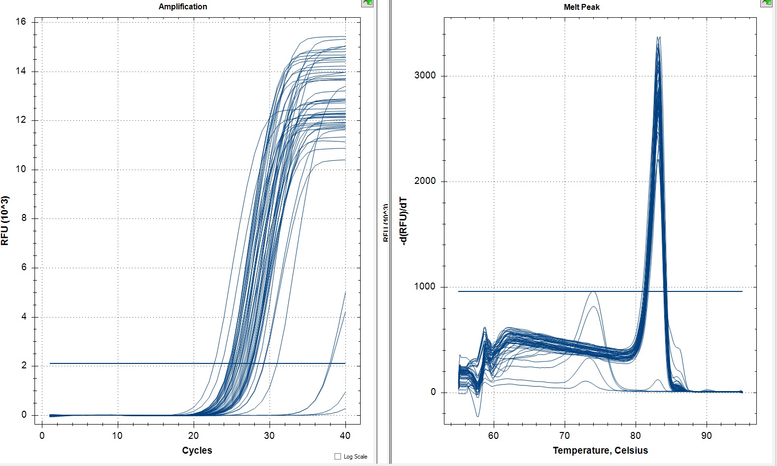
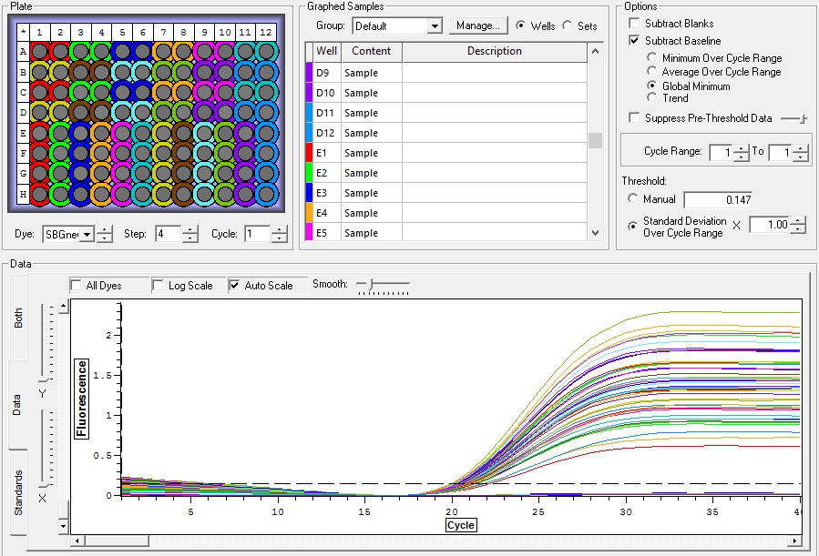
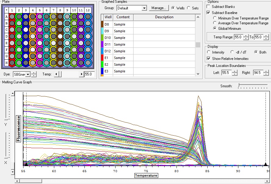
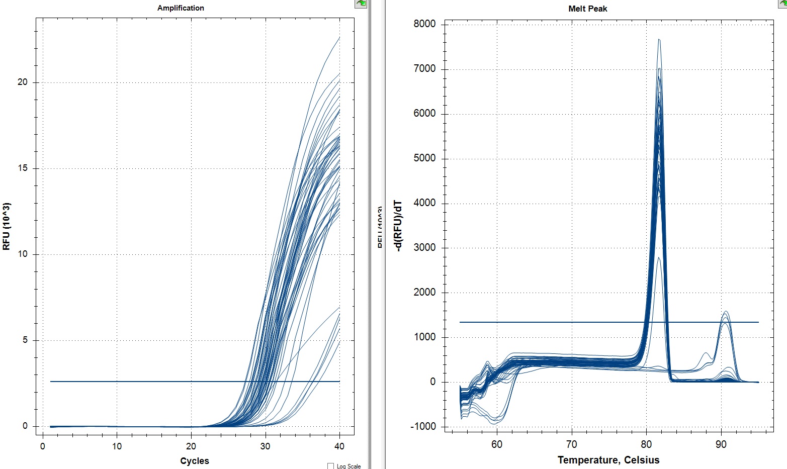
Results:
All Samples
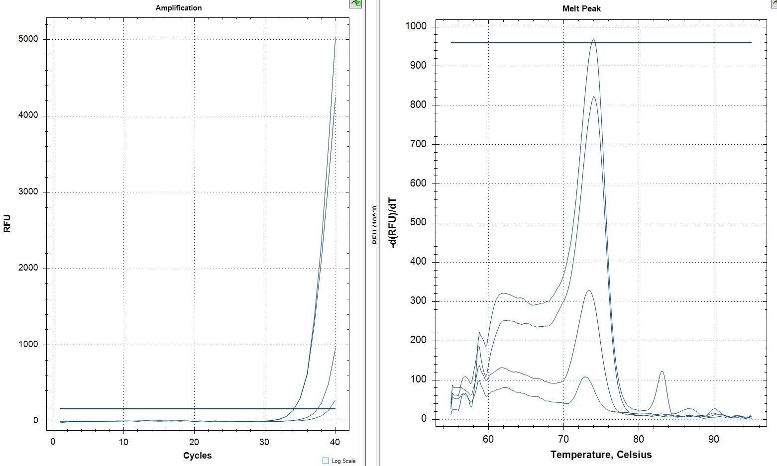
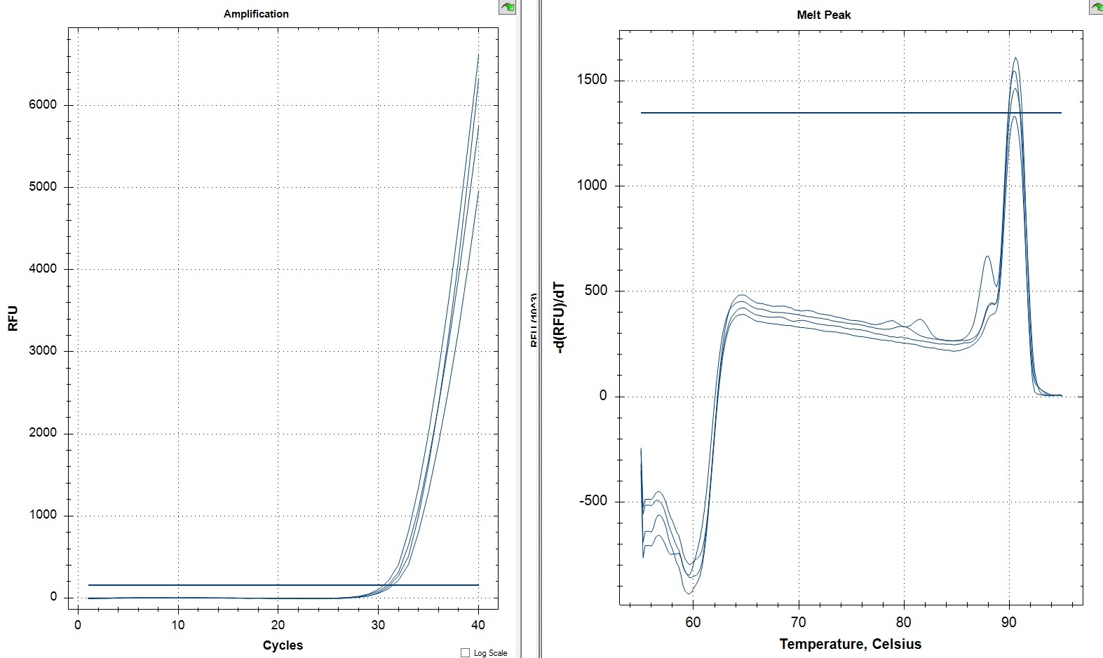
NTCs
I ran replicates of this in the future and I will post about them soon.
You can see the raw data here.
You can see the raw data here.