Tag Archives: DNA Shearing

DNA Sonication & Bioanalzyer – C. virginica gDNA for MeDIP

I transferred 8ug (136uL) of Crassotrea virginica gDNA (isolated earlier today) to two separate 1.7mL snap cap tubes for sonication/shearing.

I performed shearing at the NOAA Northwest Fisheries Science Center, using the Qsonica Q800R. Mackenzie Gavery assisted me.

Target fragment size was ~500bp.

Samples were run at the same time with the following settings:

  • 10 minutes
  • 30 seconds on, 30 seconds off
  • 25% power

After sonication, fragmentation was assessed using the Seeb Lab’s Bioanlyzer 2100 (Agilent) and the DNA 12000 Chip Kit (Agilent). NOTE: All of the reagents and the chips were past their expiration dates (most in June 2016).

Results:

Agilent 2100 Bioanalyzer Expert file (XAD): 2100 expert_DNA 12000_DE72902486_2017-12-11_13-45-31.xad

Fragmentation was successful, and pretty consistent.

Both samples appear to have an average fragment size of ~420bp. Will proceed with MeDIP, once reagents are received.

Unsheared gDNA:

DNA Isolation – C.gigas larvae from 2011 NOAA OA Experiment

DNA was isolated from the following samples:

SAMPLE ID DATE TREATMENT (ppm) # LARVAE
6B5 20110513 400 5,000
1B2 20110513 1000 5,000
6B2 20110513 400 10,000
1B1 20110513 1000 10,000
1B1 20110519 1000 NA
1B2 20110519 1000 NA
6B2 20110519 400 NA
6B1 20110519 400 NA


Some tubes contained a high quantity of algae, based on quantity of material in tube and overall green color.

Samples 1B1 & 1B2 from 20110519 have excessive quantities of algae.

Samples 6B1 & 6B1 from 20110519 have a fair amount of algae.

See pic:


Sample tubes after brief spin, prior to DNA isolation.

Prior to isolation, samples were briefly spun (12,000g, 15s @ RT). Supernatants were discarded.


DNA Isolation

DNA was isolated using the DNeasy Blood & Tissue Kit (Qiagen).

Samples were resuspended in 180uL of Buffer AL and 20uL of Proteinase K. Samples were mixed by vortexing and incubated @ 56C O/N.

The manufacturer’s protocol (Purification of Total DNA from Animal Tissues (Spin-Column Protocol)) was followed.

Due to low quantities of starting tissue, samples were eluted with 200μL of Buffer EB to maximize DNA recovery.


DNA Quantification

Samples were prepared for quantification via fluorescence using the Quant-iT DNA BR Kit (Life Technologies/Invitrogen). The manufacturer’s protocol was altered to use 5μL of sample and 5μL of standards (instead of 10μL) in each well. All samples/standards were run in duplicate and read on a FLx800 plate reader (BioTek).

Mean fluorescence of the standards were plotted with a best-fit line. The resulting equation from the best-fit line was used to determine sample concentrations from their mean fluorescence.


Results:

Calcs and resulting quantities are here:

https://docs.google.com/spreadsheets/d/1e7EF05akWeBtO7Xz0UWXhIzRlkVU5HA_rjCE3c4SPEw/edit?usp=sharing


All samples have yields great enough to proceed with shearing and bisulfite conversion.

Samples 1B1 and 1B2 from 20110519 have extremely large yields.  This is not surprising, considering the amount of algae present in the source tubes.  Will process only 500ng from each sample.


DNA Shearing

Adjusted volume of all samples to 190μL using Buffer EB (Qiagen) in 1.5mL snap-cap tubes.

Samples were sonicated/sheared in the Bioruptor (Diagenode) with the following cycling protocol:

25 cycles of:

30s on

30s off

Cycling params were adjusted from the last time I performed this, since I felt the final sheared size was a bit on the small size.

After shearing, samples were stored @ 4C until I could SpeedVac them to reduce their volumes, as the bisulfite treatment step requires volumes < 24uL.


Gel – Sheared LSU C.virginica Oil Spill gDNA (from yesterday)

Ran ~250ng of sheared C.virginica gDNA from yesterday’s shearing.

Results:

Ladder used: O’GeneRuler 100bp Ladder (ThermoFisher)

The shearing is, surprisingly, very inconsistent across the samples. The target average fragment size was ~350bp. However, most of these samples are <250bp. The MethylMiner Kit (Invitrogen) suggests that an average fragment length of 100 – 200bp is ideal for short-read high-throughput sequencing, but we’re going to perform a bisulfite conversion on these which will result in some additional fragmentation, further reducing the average fragment size. Will proceed with methylated DNA enrichment.

DNA Shearing – LSU C.virginica Oil Spill gDNA

Used the remainder of the “sheared” samples (see today’s earlier entry; ~2750ng). Brought the volumes up to 80uL and transferred to 0.5mL snap cap tubes. The volume of 80uL was selected because it’s above the minimum volume required for shearing in 0.5mL tubes (10uL according to the Biorupter 300 manual) and the MethylMiner Kit (Invitrogen) requires the input DNA volume to be <= 80uL.

DNA was sheared with the following parameters:

Low power

30 cycles of:

30s on

30s off

Target average fragment size is ~350bp.

See tomorrow (20141126) for the gel.

DNA Shearing & Size Selection – Oly Oyster gDNA RAD P1 Adapters (from 20141105)

Pooled “low quality” samples and pooled “high quality” samples separately (in 1.5mL snap cap tubes) prior to shearing to improve chances of getting similar DNA size ranges.

Samples were selected based on the gels run by Steven on Oct. 17, 2014: http://sr320.tumblr.com/

Low quality samples (5uL from each):

All rows, columns 1 -9

Higher quality samples (5uL from each):

All rows, columns 10 -12

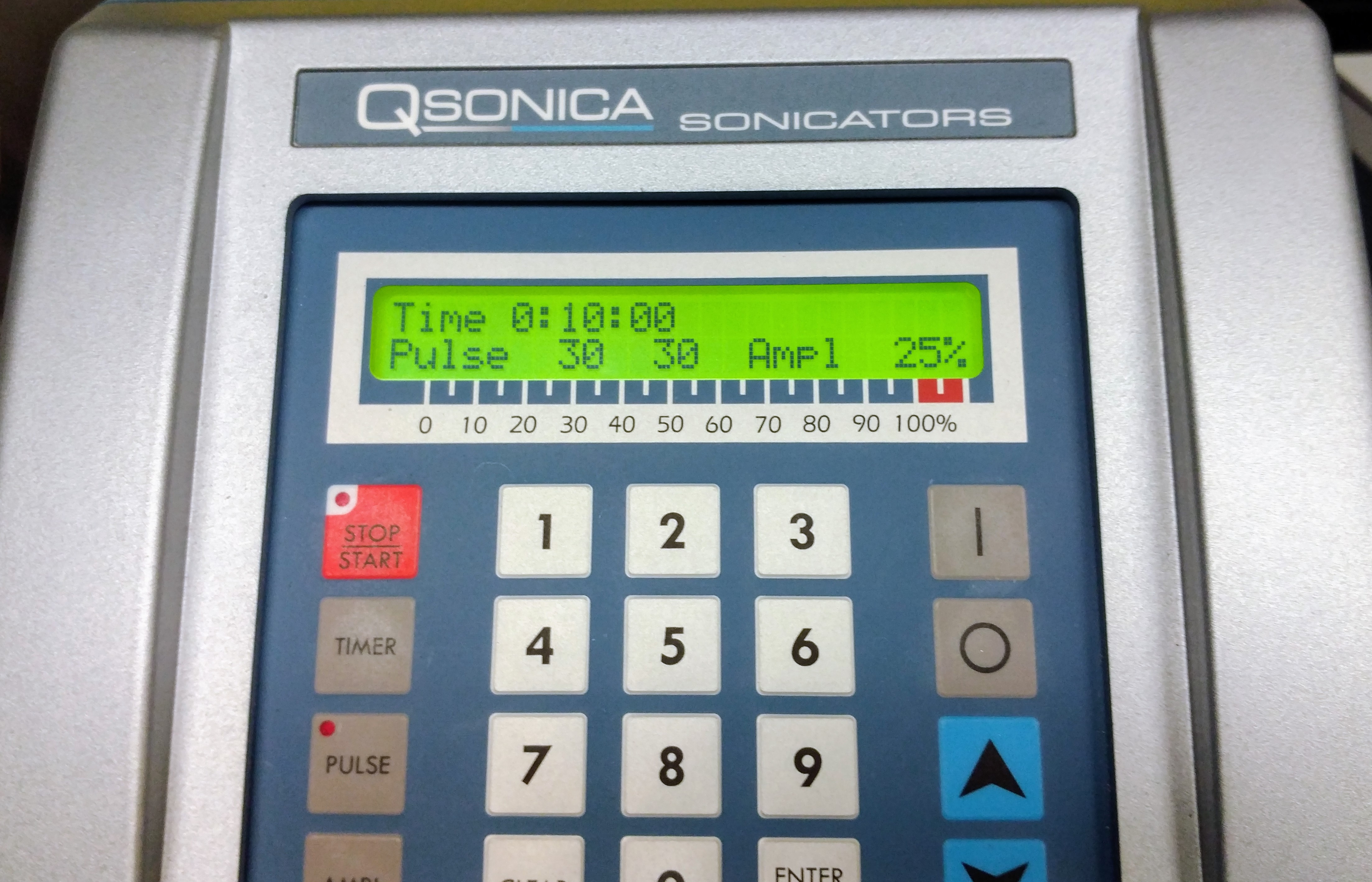
Sheared each samples with the following cycling protocols on the Biorupter Plus (Diagenode):

Low

  • 3 cycles of:
  • 30 seconds on
  • 59 seconds off


High

4 cycles of:

  • 30 seconds on
  • 59 seconds off


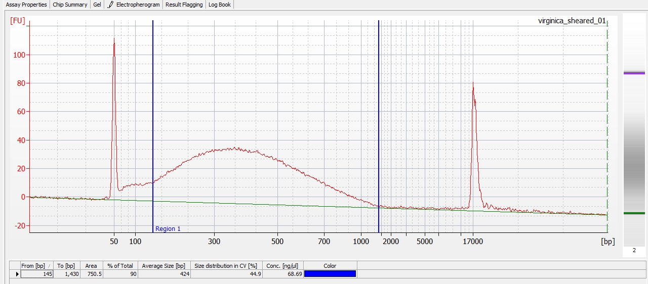
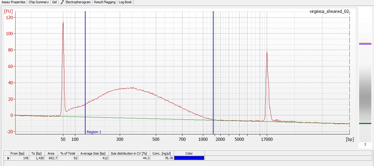
Ran a subset of sheared gDNA (5uL from each pool) on gel to verify final size range:

Gel loading:

Lane 1 – O’GeneRuler 100bp Ladder (ThermoFisher)

Lane 2 – Low quality

Lane 3 – Higher quality

I neglected to run a set of un-sheared DNA.

Both samples appear to have an average size of 200 – 400bp.

After confirming satisfactory shearing, the two samples were combined and run on a 1% agarose low TAE gel (stained with EtBr) for size selection.

O’GeneRuler 100bp Ladder (ThermoFisher)

O’GeneRuler 100bp Ladder (ThermoFisher)

Size range of sheared DNA from 300 – 500bp was excised from gel.


Gel fragment weighed 254mg.

Purified using MiniElute Gel Extraction Kit (Qiagen).

Added three volumes (762uL) of Buffer QG to gel slice.

Incubated ~10mins on rotator until gel slice was fully dissolved.

Added one gel slice volume (254uL) of isopropanol; inverted multiple times to mix.

Added 700uL to MiniElute column; spun max speed (~16,000g) 1min; discarded flow-through.

Added remainder of sample to MiniElute column; spun max speed (~16,000g) 1min; discarded flow-through.

Added 500uL of Buffer QG to MiniElute column; spun max speed (~16,000g) 1min; discarded flow-through.

Added 750uL of Buffer PE to MiniElute column; incubated @ RT for 5mins; spun max speed (~16,000g) 1min; discarded flow-through.

Spun MinElute column spun max speed (~16,000g) 1min; transferred column to clean 2.0mL tube.

Added 50uL of Buffer EB to column, incubated @ RT for 5mins and spun max speed (~16,000g) 1min; discarded column.

Stored sample @ 4C.

gDNA Sonication – SB/WB gDNA pools (prep for MeDIP) from 20100618

The previous attempt at sonication (see 20100618) failed, likely due to no using the correct equipment (tubes and Covaris adapter). The two gDNA pools, which had previously been unsuccessfully fragmented on 20100618 (SB and WB) were sonicated using a Covaris S2. Used the guidelines of the manufacturer (listed below) for shearing gDNA to a desired target size (500bp):

Duty Cycle: 5%

Intensity: 3

Cycels per Burst: 200

Time (seconds): 90

Temp (water bath): 4C

Power Mode: Frequency Sweeping

Sample Volume: 120uL

Buffer: TE

DNA Mass: ~8ug

Starting Material: >50kb

AFA Intensifier tubes and associated Covaris adapter.

After shearing, ran 250ng of each pool on a 2% TAE agarose gel for fragmentation verification.

Results:

Lane 1 – Hyperladder I

Lane 2 – R37

Lane 3 – R51

Looking at this gel, the samples have been successfully fragmented and I would estimate have generated and average fragment size of ~400bp (going from bottom to top of the Hyperladder: 200bp, 400bp, 600bp, 800bp, 1000bp). So, this looks great! Can proceed with remainder of MeDIP procedure at any time.

Additionally, I will confirm a more accurate assessment of average fragment size by running these two samples on the Agilent Bioanalyzer.

gDNA Sonication – SB/WB gDNA pools (prep for MeDIP) from earlier today

The two gDNA pools (SB and WB) were sonicated using a Covaris S2. Used the guidelines of the manufacturer (listed below) for shearing gDNA to a desired target size (500bp):

Duty Cycle: 5%

Intensity: 3

Cycels per Burst: 200

Time (seconds): 90

Temp (water bath): 4C

Power Mode: Frequency Sweeping

Sample Volume: 120uL

Buffer: TE

DNA Mass: ~8ug

Starting Material: >50kb

To be noted, the Covaris guidelines list the use of an “AFA Intensifier” tube, which I did not use (because we don’t have them).

After shearing, ran 250ng of each pool on a 2% TAE agarose gel for fragmentation verification. Also ran 250ng of pre-sonication DNA from each pool as controls.

Results:

Lane 1 – Hyperladder I

Lane 2 – R37, Un-sonicated

Lane 3 – R37, sonicated

Lane 4 – R51, Un-sonicated

Lane 5 – R51, sonicated

Sonication with the Covaris did NOT produce the desired fragmentation (500bp smear) in either sample, although the R37 sonicated samples shows a significantly greater degree of fragmentation than the R51 sonicated sample. Not sure how to explain this difference, other than the R51 sample has a greater amount of DNA. Additionally, the results could be explained by the fact that we did not use the AFA Intensifier listed in the Covaris guidelines…

Am consulting with a person in Genome Sciences who has used a Covaris for DNA fragmentation in the past to see if the AFA Intensifiers are indeed necessary and, if so, we can use two of them. Hopefully have an answer soon and be able to proceed with additional fragmentation next week.