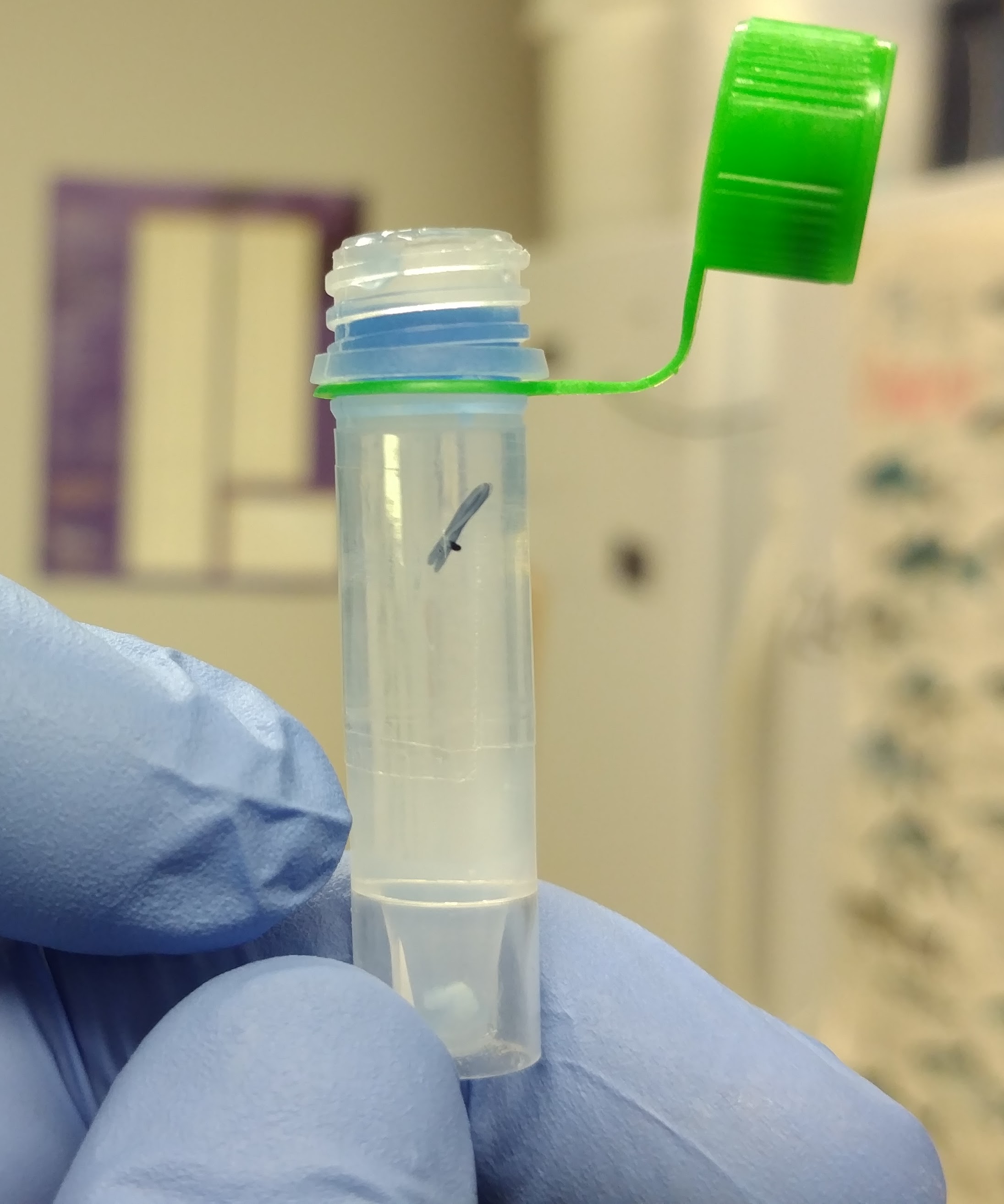
I previously isolated RNA from crab hemolymp from a lyophilized sample using TriReagent and Grace recently tried isolating RNA from crab hemolyph pellet (non-lyophilized) using TriReagent. The results for her extractions weren’t so great, so I’m giving it a shot with the following samples:
- crab 424
-
crab 429
-
crab 438
Isolated RNA using TriReagent, according to manufacturer’s protocol:
Added 1mL TriReagent to each tube, vortexed to mix/dissolve solute, incubated 5mins at RT, added 200uL of chloroform, vortexed 15s to mix, incubated at RT for 5mins, centrifuged 15mins, 12,000g, 4oC, transferred aqueous phase to new tube, added 500uL isopropanol to aqueous phase, mixed, incubated at RT for 10mins, centrifuged 8mins, 12,000g, at RT, discarded supernatant, added 1mL 75% ethanol, centrifuged 5mins, 12,000g at RT, discarded supernatant and resuspended in 10uL of 0.1% DEPC-treated H2O.
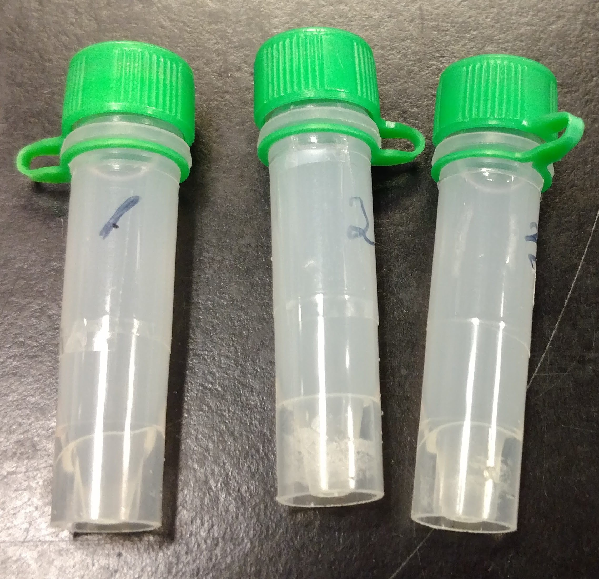
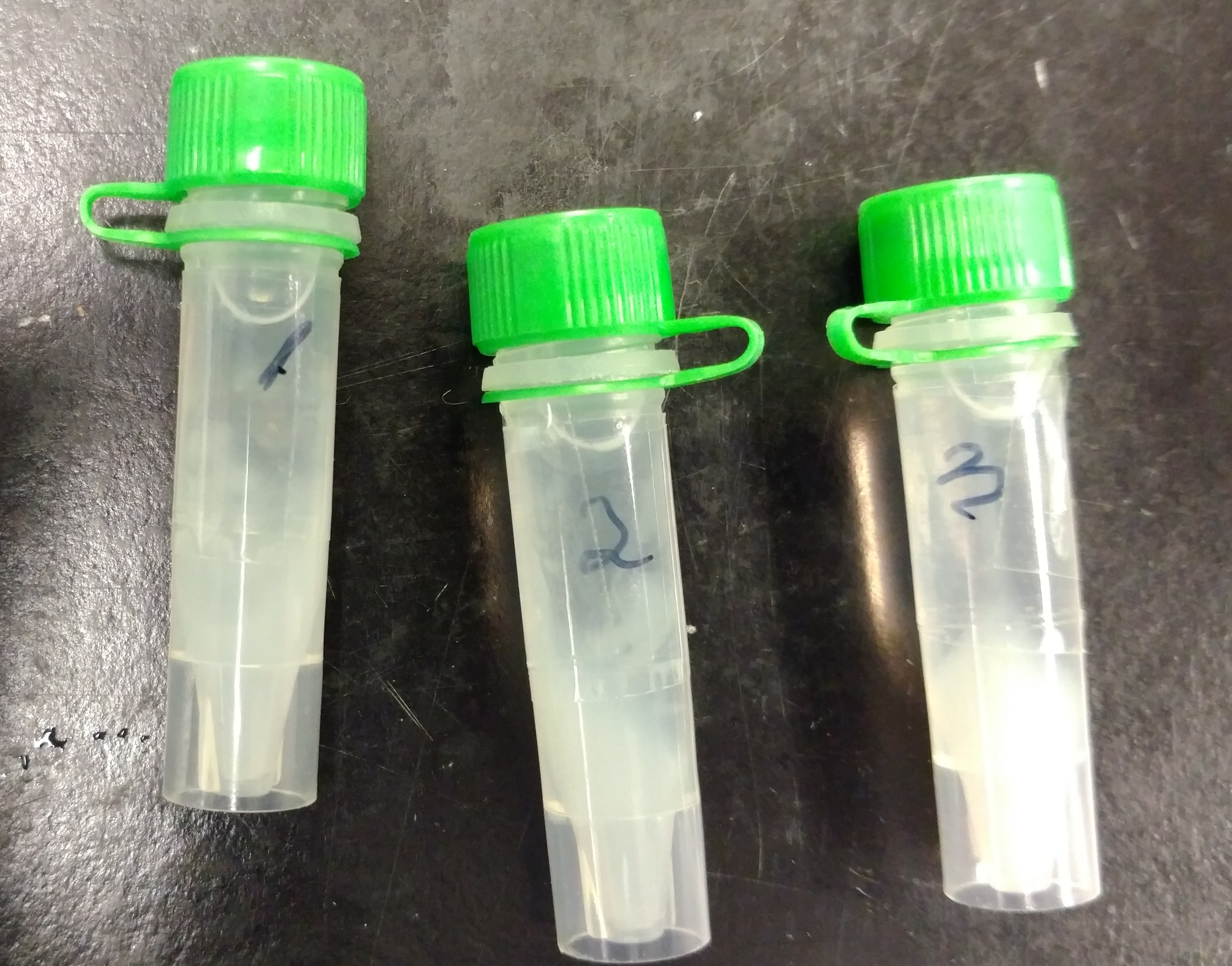
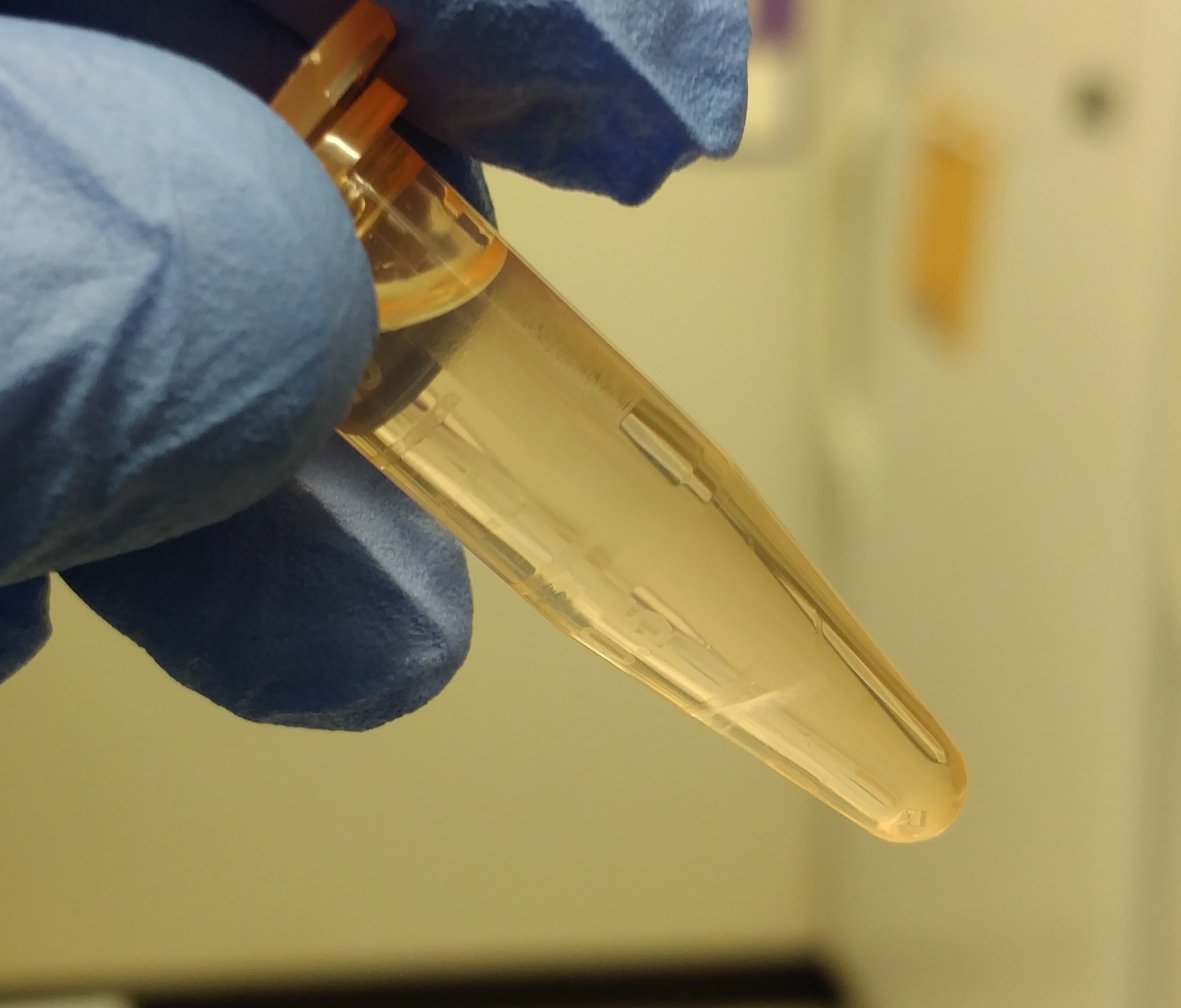
Phase separation after chloroform addition was not particularly good. Aqueous phases in sample 424 was a bit cloudy (salty?) with no defined interphase. The remaining two samples did exhibit a defined interphase and were the aqueous phases were less cloudy than sample 424, but were far from ideal.
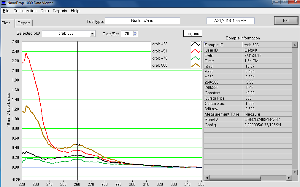
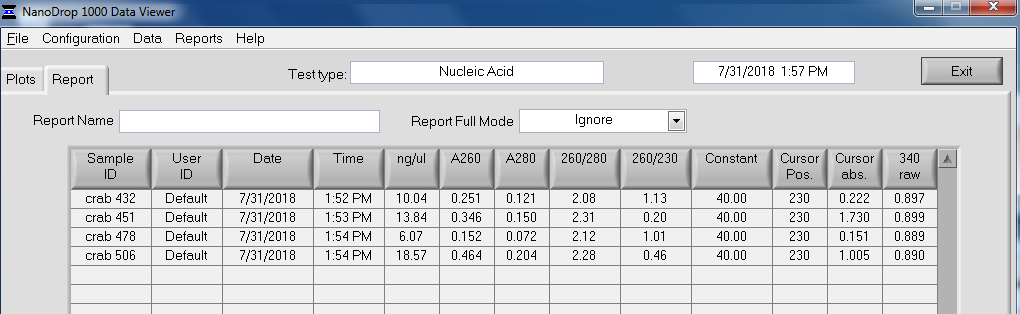
Quantified RNA using Roberts Lab Qubit 3.0 with the Qubit RNA high sensitivity kit. Used 5uL of each sample.
RESULTS
No detectable RNA in any samples. Samples were discarded.
As has been the case for all samples in this project, RNA isolation methodologies have produced wildly inconsistent results.