Tag Archives: ctenidia

Reverse Transcription – Ronit’s C.gigas DNased ctenidia RNA

Proceeded with reverse transcription of Ronit’s DNased ctenidia RNA (from 20181016).

Reverse transcription was performed using 100ng of each sample with M-MMLV Reverse Transcriptase from Promega.

Briefly, 100ng of DNased RNA was combined with oligo dT primers and brought up to a final volume of 15uL. Tubes were incubated for 5mins at 70oC in a PTC-200 thermal cycler (MJ Research), using a heated lid. Samples were immediately placed on ice.

A master mix of buffer, dNTPs, water, and M-MMLV reverse transcriptase was made, 10uL of the master mix was added to each sample, and mixed via finger flicking. Samples were incubated for 1hr at 42oC in a PTC-200 thermal cycler (MJ Research), using a heated lid, followed by a 5min incubation at 65oC.

Samples were stored on ice for use later this afternoon by Ronit.

Samples will be stored in Ronit’s -20oC box.

Reverse transcription calcs (Google Sheet):

qPCR – Ronit’s DNAsed C.gigas Ploidy/Dessication RNA with elongation factor primers

After I figured out the appropriate DNA and primers to use to detect gDNA in Crassostrea gigas samples, I checked Ronit’s DNased ctenidia RNA (from 20181016) for residual gDNA.

Elongation factor primers:

  • EF1_qPCR_5′ (SRID 309)
  • EF1_qPCR_3′ (SRID 310)

BB16 from 20090519 was used as a positive control.

Samples were run on Roberts Lab CFX Connect (BioRad). All samples were run in duplicate. See qPCR Report (Results section) for plate layout, cycling params, etc.

qPCR master mix calcs (Google Sheet):


Results

qPCR Report (PDF):

qPCR File (PCRD):

qPCR Data (CSV):

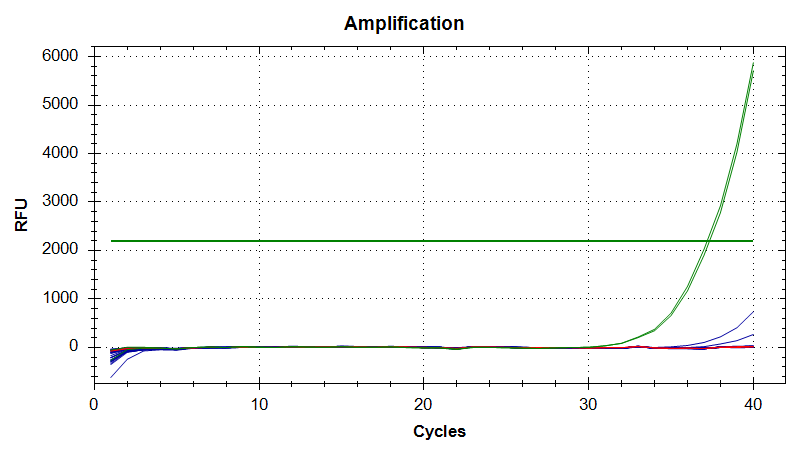
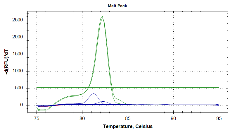
In the plots below, green is the positive control, blue are the samples, and red is the no template control (NTC).

Everything looks great! Nice, clean, gDNA-free RNA! Will proceed with reverse transcription.


Amplification Plots


Melt Curves

DNase Treatment – Ronit’s C.gigas Ploiyd/Dessication Ctenidia RNA

After quantifying Ronit’s RNA earlier today, I DNased them using the Turbo DNA-free Kit (Ambion), according to the manufacturer’s standard protocol.

Used 1000ng of RNA in a 50uL reaction in a 0.5mL thin-walled snap cap tube. Samples were mixed by finger flicking and then incubated 30mins @ 37oC in a PTC-200 thermal cylcer (MJ Research), without a heated lid.

DNase inactivation was performed (0.1 volumes of inactivation reagent; 5uL), pelleted, and supe transferred to new 1.7mL snap cap tube.

Samples were stored on ice in preparation for qPCR to test for residual gDNA.

DNase calculations are here:

Samples will be permanently stored here (Google Sheet):

RNA Quantification – Ronit’s C.gigas Ploidy/Dessication RNA

Last Friday, Ronit quantified 1:10 dilutions of the RNA I isolated on 20181003 and the RNA he finished isolating on 20181011, but two of the samples (D11-C, T10-C) were still too concentrated.

I made 1:20 dilutions (1uL RNA in 19uL 0.1% DEPC-treated H2O) and quantified them using the Roberts Lab Qubit 3.0, with the RNA HS assay. Used 1uL of the diluted RNA.


RESULTS

Qubit data (Google Sheet):

Everything looks good. Added final concentration values (Qubit data x 20, to account for dilution factor) to Ronit’s master sheet (Google Sheet):

Will proceed with DNasing.

RNA Isolation – Ronit’s C.gigas diploid/triploid dessication/heat shock ctenidia tissues

Isolated RNA from a subset of Ronit’s Crassostrea gigas ctenidia samples (see Ronit’s notebook for experiment deets):

  • D01 C

  • D02 C

  • D19 C

  • D20 C

  • T01 C

  • T02 C

  • T19 C

  • T20 C

RNA was isolated using RNAzol RT (Molecular Research Center) in the following way:

  • Unweighed, frozen tissue transferred to 500uL of RNAzol RT and immediately homogenized with disposable pestle.

  • Added additional 500uL of RNAzol RT and vortexed to mix.

  • Added 400uL of 0.1% DEPC-treated H2O, vortexed and incubated 15mins at RT.

  • Centrifuged 12,000g for 15mins at RT.

  • Transferred 750uL of supernatant to clean tube (discarded remainder), added 1 volume (750uL) of isopropanol, vortexed, and incubated at RT for 10mins.

  • Centrifuged 12,000g for 10mins at RT.

  • Discarded supernatant.

  • Washed pellet with 75% ethanol (made with 0.1% DEPC-treated H2O).

  • Centrifuged 4,000g for 2mins at RT.

  • Discarded supernatant and repeated wash steps.

Pellet was resuspended in 50uL of 0.1% DEPC-treated H2O and stored @ -80oC in Ronit’s temporary box.

DNA Isolation – Geoduck Ctenidia gDNA

Isolated additional gDNA for the genome sequencing. In an attempt to obtain better yields, I used ctenidia (instead of adductor muscle). Additionally, to try to improve the quality (260/280 & 260/230 ratios) of the gDNA, I added a chloroform step after the initial tissue homogenization.

Used 190mg of Panopea generosa ctenidia collected by Brent & Steven on 20150811.

  • Homogenized in 500μL of DNAzol.
  • Added additional 500μL of DNAzol.
  • Centrifuged 12,000g, 10mins, @ RT.
  • Split supernatant equally into two tubes.
  • Added 500μL of chloroform and mixed moderately fast by hand.
  • Centrifuged 12,000g, 10mins, RT.
  • Combined aqueous phases from both tubes in a clean tube.
  • Added 500μL of 100% EtOH and mixed by inversion.
  • Spooled precipitated gDNA and transferred to clean tube.
  • Performed 3 washes w/70% EtOH.
  • Dried pellet 3mins.
  • Resuspended in 200μL of Buffer EB (Qiagen).
  • Centrifuged 10,000g, 5mins, RT to pellet insoluble material.
  • Transferred supe to clean tube.

DNA was quantified using two methods: NanoDrop1000 & Qubit 3.0 (ThermoFisher).

For the Qubit, the samples were quantified using the Qubit dsDNA BR reagents (Invitrogen) according to the manufacturer’s protocol and used 1μL of sample for measurement.

Results:

Qubit Data (Google Sheet): 20151125_qubit_gDNA_geoduck_oly_quants

METHOD CONCENTRATION (ng/μL) TOTAL (μg)
Qubit 105 21.0
NanoDrop1000 173 34.6


Yield is definitely much, much better than adductor muscle! Should’ve switched to a different tissue a long time ago! We should finally have sufficient quantities of gDNA to allow for BGI to proceed with the rest of the genome sequencing! Will run sample on gel to evaluate integrity and then send off to BGI.

The NanoDrop & Qubit numbers still aren’t close (as expected).

The addition of the chloroform step definitely helped improve the 260/280 OD ratio (see below). However, the addition of that step had no noticeable impact on the 260/230 OD ratios, which is a bit disappointing.


NanoDrop Absorbance Values & Plots


Oyster Sampling – Oly Fidalgo 2SN, 2HL, 2NF Reciprocal Transplants Final Samplings

The remaining Olympia oysters from Jake Heare’s reciprocal transplant experiment have been retrieved from field sites and are awaiting sampling. The oysters have been stored in the cold room (temp?) for 15 days so far.

The previous sampling scheme was described here: DNA Isolations – Fidalgo 2SN Reciprocal Transplants Final Samplings

Sampling scheme for today was as follows:

  1. Assign unique number to oysters (1-100 for each of the three populations)
  2. Photograph with ruler for future shell measurements
  3. Weigh oysters
  4. Dissect ctenidia for DNA isolation in 350μL MBL1 Buffer + 25μL Proteinase K (reagents part of the E.Z.N.A. Mollusc Kit [Omega BioTek)
  5. Preserve portion of remaining body tissue (not viscera; gonad/digestive gland) in 1mL RNAlater (Life Technologies)

Ctenidia samples were stored -80C in the buffer/pro k solution for DNA isolation at a later date.

RNAlater samples will be stored over the weekend at 4C and then transferred to -20C for long term storage.

All oyster data is here (Google Sheet): Oly reciprocal final sampling

All photos from today’s sampling are here: Oyster Measurement Photos

DNA Isolations – Fidalgo 2SN Reciprocal Transplants Final Samplings

The remaining Olympia oysters from Jake Heare’s reciprocal transplant experiment have been retrieved from field sites and are awaiting sampling. The oysters have been stored in the cold room (temp?) for 6 days so far.

Sampling scheme is as follows:

  1. Assign unique number to oysters
  2. Photograph with ruler for future shell measurements
  3. Weigh oysters
  4. Dissect ctenidia for DNA isolation
  5. Dissect & discard viscera (e.g. digestive gland and gonad)
  6. Weigh remaining body
  7. Preserve remaining body in RNAlater
  8. Weigh empty shells


Mrunmayee photographed & initiated dissections of oysters #3 – 8
. I took over for oyster #9 -14.

All oyster data is here (Google Sheet): Oly reciprocal final sampling

DNA was isolated using the E.Z.N.A. Mollusc Kit (Omega Biotek) according to the manufacturer’s protocol with the following changes:

  • No optional steps were performed
  • Ctenidia tissue was lysed for 3hrs @ 60C
  • Single elution of 50μL

Samples were stored @ -20C in: Oly gDNA Oly Reciprocal Transplant Final Sampling Box #1.

Reverse Transcription – O.lurida DNased RNA 1hr post-mechanical stress

Performed reverse transcription on the Olympia oyster DNased RNA from the 1hr post-mechanical stress samples from Jake’s project. To accommodate the large numbers of anticipated genes to be targeted in subsequent qPCRs, I prepared 100μL reactions (normally, 25μL reactions are prepared) using 250ng of each DNased RNA. A 1:10 dilution of the oligo dT primers (Promega) was prepared to improve pipetting accuracy. All incubations were performed in a thermal cycler without using a heated lid.

DNased RNA was combined with NanoPure H2O and oligo dT primers in 24 wells of a PCR plate, heated @ 70C for 10mins and immediately placed on ice. After 5mins, the plate was spun 2000g @ RT for 2mins and returned to ice.

25.25μL of a master mix containing 5x M-MLV Buffer (Promega), dNTPs (10mM each; Promega), and M-MLV Reverse Transcriptase (50U/rxn; Promega) was distributed to each well and mixed via pipetting. The plate was heated @ 42C for 1hr, 95C for 3mins. The plate was spun 2000g @ RT for 2mins and then stored @ -20C.

Plate layout and all calculations can be found here (Google Sheet): 20150806_Jake_oly_mech_stress_cDNA_calcs