Tag Archives: RNAlater

RNA Isolation – Lyophilized Tanner Crab Hemolymph in RNAlater

Due to difficulties getting RNA from hemolymph samples stored in RNAlater, Grace is testing out lyophilizing samples before extraction. Who knows what impact this will have on RNA, but it’s worth a shot!

Isolated RNA from three crab hemolymph samples preserved in RNAlater (Test 1, Test 2, Test 3) that had been lyophilized overnight last week.

Samples were provided by Grace.

I believe the primary purpose for this particular test was to verify that the freeze dryer was a feasible tool, since Grace experienced a minor mishap when she attempted the lyohpilization initially.

Lyophilization was successful, without any mess.

TEST 3 LYOPHILIZATION
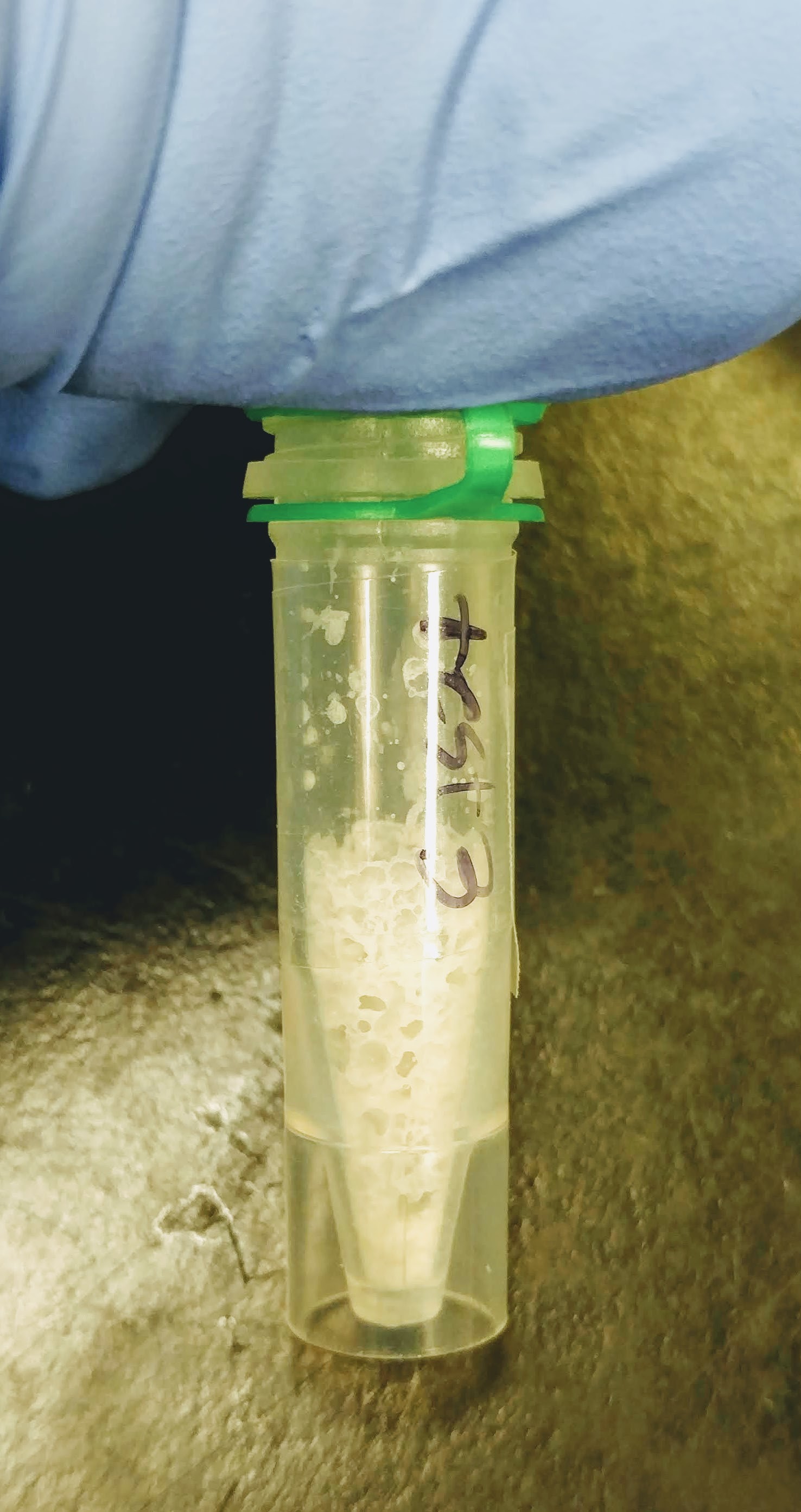

Isolated RNA using TriReagent, according to manufacturer’s protocol:

Added 1mL TriReagent to each tube, vortexed to mix/dissolve solute, incubated 5mins at RT, added 200uL of chloroform, vortexed 15s to mix, incubated at RT for 5mins, centrifuged 15mins, 12,000g, 4oC, transferred aqueous phase to new tube, added 500uL isopropanol to aqueous phase, mixed, incubated at RT for 10mins, centrifuged 8mins, 12,000g, at RT, discarded supernatant, added 1mL 75% ethanol, centrifuged 5mins, 12,000g at RT, discarded supernatant and resuspended in 10uL of 0.1% DEPC-treated H2O.

Quantified RNA using Roberts Lab Qubit 3.0 with the Qubit RNA high sensitivity kit. Used 5uL of each sample.


RESULTS

Qubit (Google Sheet):

Only one sample (Test 3) had detectable levels of RNA (20.4ng/uL).

So, this little test demonstrates that RNA can be isolated from lyophilized samples and extracted with TriReagent. However, I have not evaluated RNA integrity on the Bioanalyzer. I think Grace has some additional samples she wanted to test this method on, so I think we’ll wait until there are more samples before we use the Bioanalyzer.

Will give sample to Grace for -80oC storage.
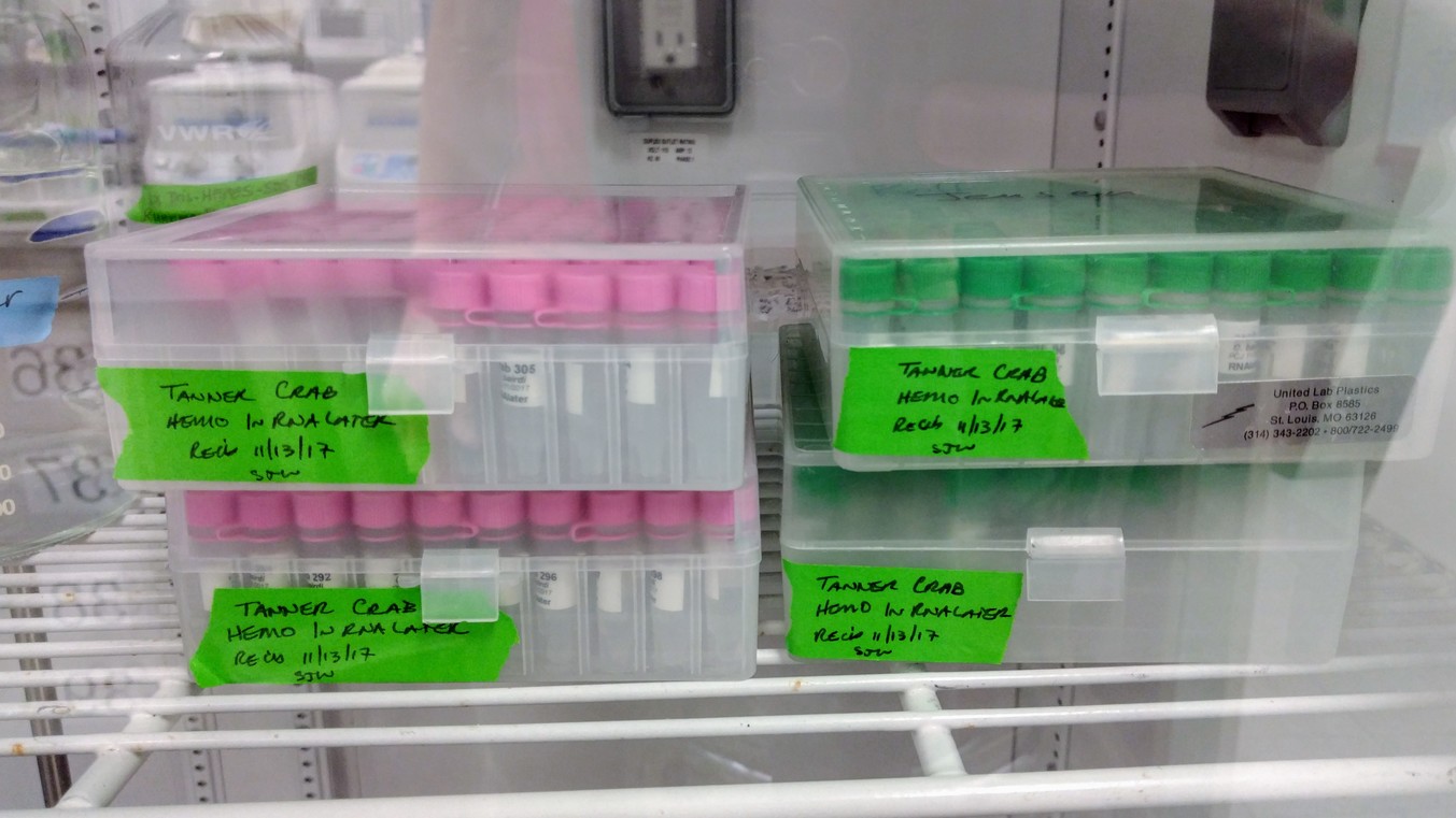
Samples Received – C.gigas Tissue & DNA from Katie Lotterhos

Received 6 samples from Katie today. The box was labeled and stored @ -20C.


Here description of the samples, via email:

Lotterhos samples (gigas) arriving tomorrow

1) Mantle tissue samples of C. gigas were collected on 20140705 (source: Pipestem Inlet) by KEL
2) Extraction on 20141028 by VG using Qiagen DNAeasy Blood and Tissue Kit
3) Beadwash on 20150720 by VG using homemade sera-mag speed beads
4) Qubit 3.0 quantification on 20151206 by KEL and the following amounts were sent:

M1: 13 uL of 386 ug/mL
M2: 13.8 uL of 326 ug/mL
M3: 13.15 uL of 380 ug/mL (solution looked cloudy)


Oyster Sampling – Oly Fidalgo 2SN, 2HL, 2NF Reciprocal Transplants Final Samplings

The remaining Olympia oysters from Jake Heare’s reciprocal transplant experiment have been retrieved from field sites and are awaiting sampling. The oysters have been stored in the cold room (temp?) for 15 days so far.

The previous sampling scheme was described here: DNA Isolations – Fidalgo 2SN Reciprocal Transplants Final Samplings

Sampling scheme for today was as follows:

  1. Assign unique number to oysters (1-100 for each of the three populations)
  2. Photograph with ruler for future shell measurements
  3. Weigh oysters
  4. Dissect ctenidia for DNA isolation in 350μL MBL1 Buffer + 25μL Proteinase K (reagents part of the E.Z.N.A. Mollusc Kit [Omega BioTek)
  5. Preserve portion of remaining body tissue (not viscera; gonad/digestive gland) in 1mL RNAlater (Life Technologies)

Ctenidia samples were stored -80C in the buffer/pro k solution for DNA isolation at a later date.

RNAlater samples will be stored over the weekend at 4C and then transferred to -20C for long term storage.

All oyster data is here (Google Sheet): Oly reciprocal final sampling

All photos from today’s sampling are here: Oyster Measurement Photos

DNA Isolations – Fidalgo 2SN Reciprocal Transplants Final Samplings

The remaining Olympia oysters from Jake Heare’s reciprocal transplant experiment have been retrieved from field sites and are awaiting sampling. The oysters have been stored in the cold room (temp?) for 6 days so far.

Sampling scheme is as follows:

  1. Assign unique number to oysters
  2. Photograph with ruler for future shell measurements
  3. Weigh oysters
  4. Dissect ctenidia for DNA isolation
  5. Dissect & discard viscera (e.g. digestive gland and gonad)
  6. Weigh remaining body
  7. Preserve remaining body in RNAlater
  8. Weigh empty shells


Mrunmayee photographed & initiated dissections of oysters #3 – 8
. I took over for oyster #9 -14.

All oyster data is here (Google Sheet): Oly reciprocal final sampling

DNA was isolated using the E.Z.N.A. Mollusc Kit (Omega Biotek) according to the manufacturer’s protocol with the following changes:

  • No optional steps were performed
  • Ctenidia tissue was lysed for 3hrs @ 60C
  • Single elution of 50μL

Samples were stored @ -20C in: Oly gDNA Oly Reciprocal Transplant Final Sampling Box #1.

RNA Isolation – Jessica’s Geoduck Larval Stages

Isolated RNA from the following samples provided by Jessica Blanchette (stored in RNA later):

  • Trocophore 1 (T1)
  • Trocophore 2 (T2)
  • Veliger 1 (V1)
  • Veliger 2 (V2)
  • Settlers Interphase 1 (S1)
  • Settlers Interphase 2 (S2)

The tocophore and veliger larval stages are neutrally bouyant (i.e. will not pellet when centrifuged). In order to separate them from the RNA Later, I used a fine mesh (don’t know mesh size; bag was labeled “Unknown”) as a “guard” between the pipette tip and the larvae. Removed RNA Later from those two groups in this fashion. However, a significant portion of the larvae in these tubes adhered to the outside of the mesh. I left the mesh “guard” in the tube, added 1mL of TriReagent and vortexed. The mesh quickly dissolved in the TriReagent, creating a milky white mix.

For the settlers samples, there was a such a large pellet already in the existing tubes, I just took ~75uL of this material, transferred to a clean tube and added 1mL of TriReagent. However, most of the debris that I transferred dissolved extremely quickly. I was expecting there to more insoluble “debris”, because marine bivalve larval shells generally don’t readily dissolve in the presence of TriReagent. So, I suspect that much of the settlers samples is not really geoduck larvae.

Due to time constraints, stored all samples O/N @-80C in TriReagent.

Samples were thawed and RNA was isolated, and DNased, using the Direct-zol RNA Miniprep Kit (ZymoResearch), eluted with 50uL of 0.1% DEPC-treated H2O, and spec’d on the NanoDrop1000.

Prior to isolation, sample V1 showed a clear phase separation that none of the other samples exhibited. Sample V1 had a pink, goopy layer on top of a clear, low-viscosity layer. All other samples retained the uniform pink coloration imparted by the TriReagent. Additionally, after addition of the EtOH in the procedure to sample V1, a large amount of white precipitate formed and settled to the bottom of the tube. This did not happen in any other samples.

Samples were stored @ -80C in “Shellfish RNA Box #5

Results:

Overall, the yields are relatively low, as expected. Virtually all of the samples have poor OD260/280 values. Although not shown, there was a consistent shift in peak absorbance from 260nm towards 270nm, leading to the poor OD260/280 values.

RNA Isolation – Colleens’ Sea Star Coelomycetes Samples

Isolated RNA from the following samples stored in RNAlater:

  • TH52 3.28.14 c-fluid
  • TH54 3.28.14 c-fluid
  • CH55 3.28.14 c-fluid
  • CH56 3.28.14 c-fluid
  • CH57 3.28.14 c-fluid
  • TH65 3.28.14 c-fluid
  • TH66 3.28.14 c-fluid
  • TH67 3.28.14 c-fluid

Spun samples 5000g, 20mins @ RT to pellet any cells. Discarded supe. Resuspended cells/debris in 1mL TriReagent. Disrupted cells by pipetting and vortexting. RNA was isolated using the Direct-zol RNA Miniprep Kit (ZymoResearch). RNA was DNase treated on-column, as described in the manufacturer’s protocol, using DNase I. RNA was eluted from the columns using 25uL of nuclease-free H2O and spec’d on a NanoDrop1000.

Results:

So, this is disheartening. Overall, the RNA looks pretty crappy; poor 260/280 ratios and a general shift in absorbance to 270nm. Plus, the yields aren’t that great. Maybe RNA left on the column and/or some sort of contaminant pushing these readings out of whack?

I will perform another elution on the columns with 50uL of nuclease-free H2O and spec that elution set:

There’s still a shift in the peak absorbance in most samples to 270nm… I’m going to combine the two sets of elutions and spec:

Although the 260/280 values are significantly better, there’s still this persistent shift of peak absorbance to 270nm. I contacted technical support for the kit and they say the absorbance shift is indicative of phenol contamination. They have advised that I add a volume of TriReagent to the RNA and re-run it through a new set of columns, following the entire RNA isolation protocol.

RNA Isolation – Sea Star Coelomocytes (provided by Colleen Burge)

Tried another method of RNA Isolation for comparison with regular TriReagent method.

Used the Direct-zol RNA MiniPrep Kit (Zymo Research) on the following samples stored in RNAlater:

  • P6 Control
  • P16 Filt. Inj.

Pelleted samples in RNAlater by spinning 5000g, 10mins @ RT. Removed RNAlater, lysed pellets in 1mL TriReagent. Split each sample equally into two tubes (500uL in each tube). Added equal volumes of 100% ethanol to each tube and vortexed. Transferred samples to spin columns and followe manufacturer’s protocol. Eluted with 25uL of nuclease-free H2O (provided in kit). Spec’d on NanoDrop1000.

Results:

RNA quality is very good (based on 260/280 ratios). This turned out much better than the previous attmpt using the basic TriReagent method. However, the previous attempt (see 20140401) may have been compromised by me being too aggressive when collecting the aqueous phase. Knowing how little sample was present, I may have been overzealous in trying to gather too much of the aqueous phase, leading to the phenol carryover that was evident.

Regardless, these columns seem to do an excellent job of eliminating even salt carryover, as we frequently see high absorbance at 230nm with marine samples; particularly those stored in RNAlater.

RNA Isolation – Sea Star Coelomocytes (provided by Colleen Burge)

Isolated RNA from the following samples (stored in RNAlater):

P18 Control 3/17/14

P10 Filt. Inj. 3/17/14

These were “trial” RNA isolation runs to determine what yields we could expect from samples of this nature.

Both samples had very small tissue/cell pellets. Tubes were spun @ 5000g for 10mins at RT to ensure all cells were pelleted. RNAlater was removed and pellets were lysed using 1000uL of TriReagent, supplemented with 8uL of PolyAcryl carrier. PolyAcryl Carrier was used to enhance RNA recovery from such small starting materials. Remainder of procedure followed manufacturer’s protocol. RNA was resuspended in 20uL of 0.1% DEPC-H2O and spec’d on a NandoDrop1000.

Results:

As can be seen by the absorbance spectrum plots (top image), there is clear phenol contamination (indicated by shift of absorbance peak to 270nm, instead of the peak being at 260nm). Additionally, there’re large peaks at 230nm in each of the two samples, suggesting other contamination (high residual salts, ethanol?). Additionally, the 260/280 ratios are subpar for RNA quality (i.e. <1.9). However, these ratios could be skewed by the the residual phenol present in both samples. I may perform an ethanol precipitation on these just to see if I can get them cleaned up.

Yields for both samples are very promising.

RNA Isolation – C.gigas Larvae from Taylor Summer 2011

Samples had been stored in RNA Later (Ambion). Samples were pelleted and the RNA Later supe removed. Samples were washed (2x) with 1mL TE (pH = 8.0) to remove excess salt resulting from the RNA Later. Samples were split, roughly equally, into two separate tubes. Samples were pelleted and the supe removed. One tube from each sample was set aside for gDNA isolation using DNAzol (MRC). The other tube was vortexed vigorously in TriReagent (MRC) and the then treated according to protocol. Samples were resuspended in 100uL of 0.1% DEPC-H2O and spec’d on the Roberts Lab NanoDrop 1000.

Results:

Overall, the samples look really good. Some samples (280, 434 & 605) required re-specing after the NanoDrop was reblanked in order to get a reading without an error message. They will be DNased and then reverse transcribed.