Tag Archives: hemolymph

Data Received – Chionoecetes bairdi RNAseq & FastQC Analysis

We received Grace’s 100bp PE NovaSeq (Illumian) RNAseq data from the Northwest Genomics Center today.

Data was downloaded via their Aspera browser plugin and rsynced to:

MD5 checksums were generated (md5sum on Ubuntu):


321ec408ba7e0f0be1929ca44871f963  304428_S1_L001_R1_001.fastq.gz
b95c69f755c9c42d9203429119d4234d  304428_S1_L001_R2_001.fastq.gz
a0fd8db312057dedd480231d4d125fd3  304428_S1_L002_R1_001.fastq.gz
c6e70ef7f3c8a866851a1b9453aef36a  304428_S1_L002_R2_001.fastq.gz

FastQC analysis was run, followed by MultiQC.

Output folder (gannet/Atumefaciens):

MultiQC Report (HTML):

Nightingales spreadsheet was updated with file info and FastQC info:

RNA Isolation – Lyophilized Tanner Crab Hemolymph in RNAlater

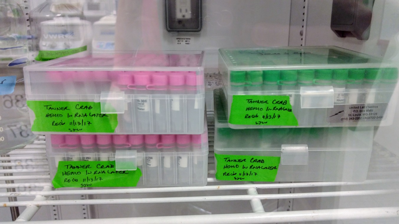
Due to difficulties getting RNA from hemolymph samples stored in RNAlater, Grace is testing out lyophilizing samples before extraction. Who knows what impact this will have on RNA, but it’s worth a shot!

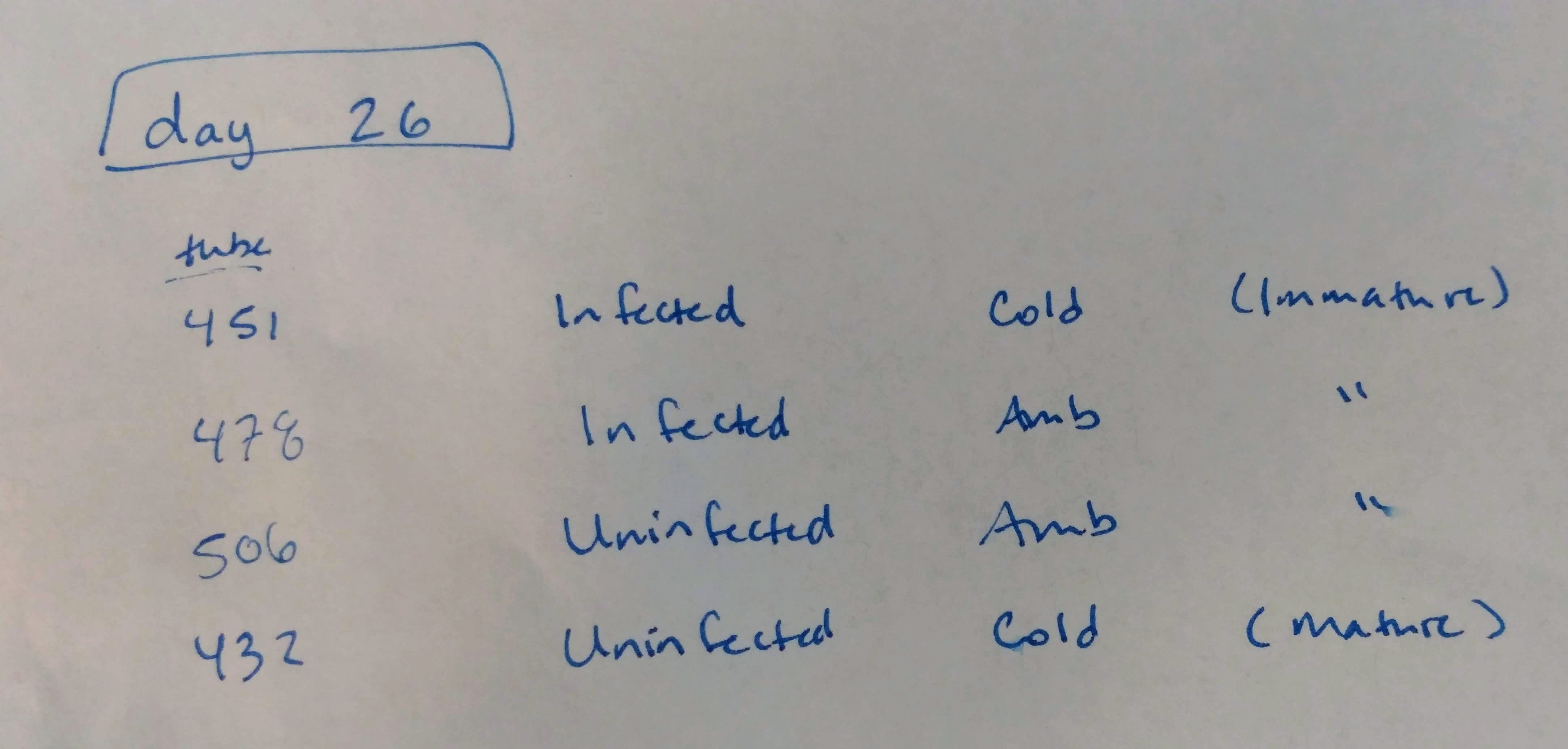
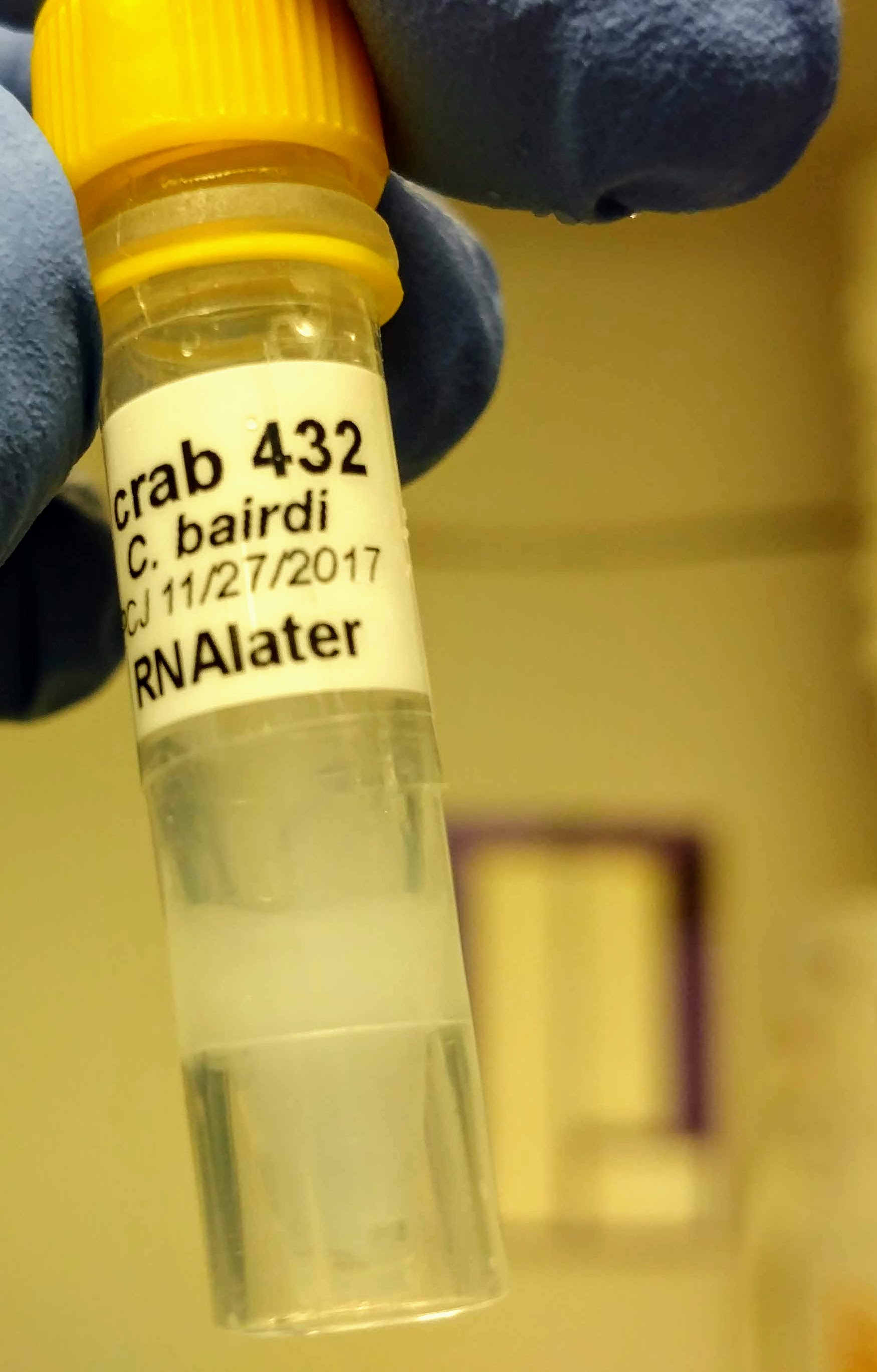
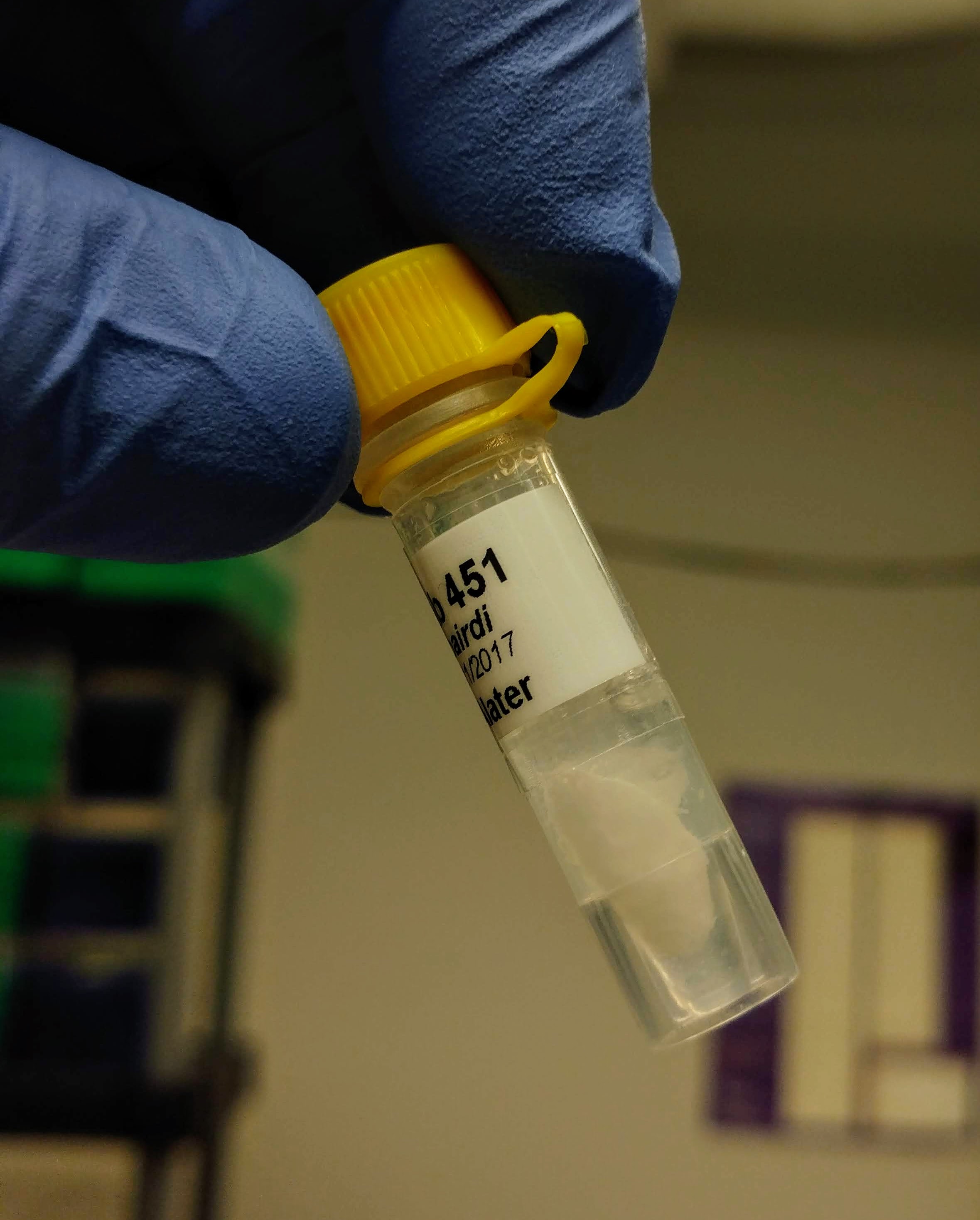
Isolated RNA from three crab hemolymph samples preserved in RNAlater (Test 1, Test 2, Test 3) that had been lyophilized overnight last week.

Samples were provided by Grace.

I believe the primary purpose for this particular test was to verify that the freeze dryer was a feasible tool, since Grace experienced a minor mishap when she attempted the lyohpilization initially.

Lyophilization was successful, without any mess.

TEST 3 LYOPHILIZATION


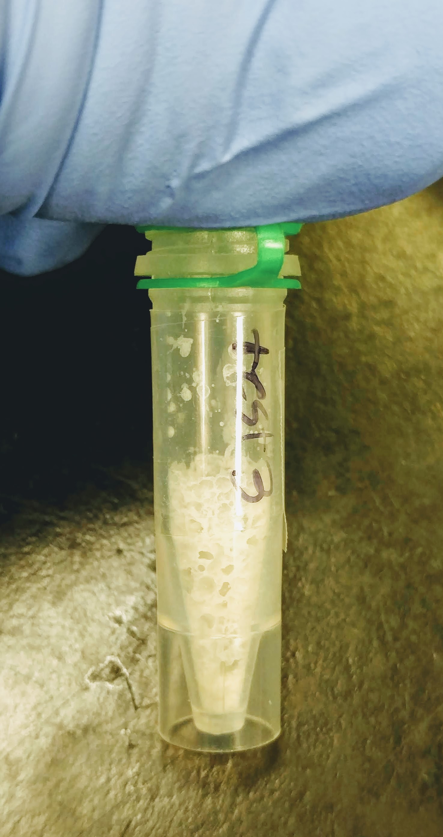
Isolated RNA using TriReagent, according to manufacturer’s protocol:

Added 1mL TriReagent to each tube, vortexed to mix/dissolve solute, incubated 5mins at RT, added 200uL of chloroform, vortexed 15s to mix, incubated at RT for 5mins, centrifuged 15mins, 12,000g, 4oC, transferred aqueous phase to new tube, added 500uL isopropanol to aqueous phase, mixed, incubated at RT for 10mins, centrifuged 8mins, 12,000g, at RT, discarded supernatant, added 1mL 75% ethanol, centrifuged 5mins, 12,000g at RT, discarded supernatant and resuspended in 10uL of 0.1% DEPC-treated H2O.

Quantified RNA using Roberts Lab Qubit 3.0 with the Qubit RNA high sensitivity kit. Used 5uL of each sample.
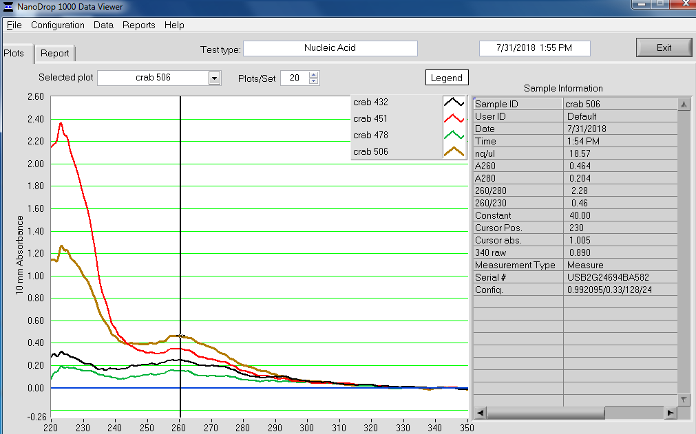
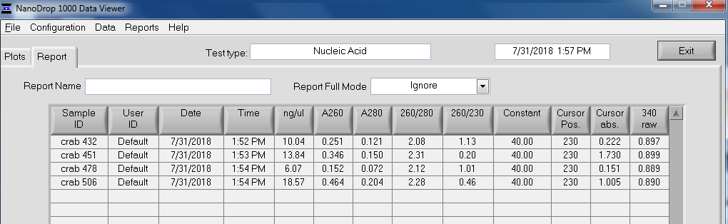

RESULTS

Qubit (Google Sheet):

Only one sample (Test 3) had detectable levels of RNA (20.4ng/uL).

So, this little test demonstrates that RNA can be isolated from lyophilized samples and extracted with TriReagent. However, I have not evaluated RNA integrity on the Bioanalyzer. I think Grace has some additional samples she wanted to test this method on, so I think we’ll wait until there are more samples before we use the Bioanalyzer.

Will give sample to Grace for -80oC storage.

RNA Isolation & Quantificaiton – Tanner Crab Hemolymph

Isolated RNA from 40 Tanner crab hemolymph samples selected by Grace with the RNeasy Plus Micro Kit (Qiagen) according to the manufacturer’s protocol, with the following modifications:

  • Added mercaptoethanol (2-ME) to Buffer RLT Plus.

  • All spins were at 21,130g

  • Did not add RNA carrier

  • Used QIAshredder columns to aid in homogenization and removal of insoluble material

  • Eluted with 14uL

RNA was quantified using the Qubit RNA HS (high sensitivity) Assay and run on the Roberts Lab Qubit 3.0.

Used 1uL of sample for quantification.

RNA was returned to the -80C box from where original samples had been stored (Rack 2, Row 3, Column 4).


RESULTS

Qubit quantification (Google Sheet):

Overall, the results aren’t great. Only 15 samples (out of 40) had detectable amounts of RNA. Yields from those 15 samples ranged from 40ng – 300ng, with most landing between 50 – 100ng.

Will pass info along to Grace. Will likely meet with her and Steven to discuss plan on how to move forward.

RNA Isolation – Tanner Crab Hemolymph Using RNeasy Plus Mini Kit

Tanner crab RNA has proved a bit troublesome. As such, Steven asked me to try isolating some RNA using the RNeasy Plus Mini Kit (Qiagen) to see how things would turn out.

Grace provided me with the following samples:


Crab hemolymph had been collected (100uL?) and preserved with 1mL (?) of RNAlater. Grace pelleted the samples, removed the supernatant, and stored the pelleted material at -80C. Here’s what that looked like:


RNA was isolated according to the manufacturer’s protocol – following guideline for samples with < 1 x 10⁶ cells.

One interesting thing that happened is a precipitate formed after adding the initial buffer to the sample:

A solid precipitate formed in each of the tubes that could not be dispersed – it actually looked like a small piece of paper was now present in each tube.

Samples were spun and the supernatant was utilized (this was the normal progression of the protocol, regardless of this precipitate forming).

Samples were eluted with 30uL of nuclease-free water.

Samples were quantified using the Roberts Lab Qubit 3.0 with the RNA High Sensitivity asssay (Invitrogen). Used 5uL of sample for measurements.

Samples were also assessed with the Roberts Lab NandoDrop1000. Samples were recovered from the pedestal after measurement.

RNA was given to Grace for storage at -80C.


RESULTS

Qubit measurements (Google Sheet):
20180731_qubit_RNA_crab_isos


NanoDrop Spec Curves:


NanoDrop Table:


Overall, the isolation looks pretty good. The purity looks good (NanoDrop 260/280 ratios) and the absorbance peak at 260nm is exactly where we would want/expect it to be.

The yields (according to the Qubit) are OK. They range from ~37ng – 350ng.

The important part is that this method produced clean RNA, which means the quantification is believable. I think Grace’s earlier RNA isolations using RNAzol RT had too much contamination carried over, leading to incorrect quantification measurements.

Going forward, I think we need to use some sort of isolation kit, however, we will be testing out good, old TriReagent as well.

Received Hard Clam Samples from Rutgers

30 gill tissue samples in RNA Later from CA, MA, & MAX each.

30 hemolymph samples (in RNA Later?) from CA, MA, & MAX each.

Presumably these are from the same individuals. Tubes were boxed (a total of 3 boxes), labeled and stored @ -80C.

Here is a note included from Emily with the samples .

Package – Hard Clam Samples from Rutgers

Rec’d package of hard clam samples from Emily @ Rutgers on wet ice. Package contained numerous 1.5mL snap cap tubes separated in to groups in zip lock bags. Stored temporarily @ 4C. Will catalog and then store @ -80C.

Three documents included with package:

Note from Emily

Sample info, pg. #1

Sample info, pg. #2

Samples Received – Hard Clam samples from Rutgers and MBL

 *Important Note: These were received while I was out of lab. This notebook entry was added 20101021*

Received sets of gill tissue and hemolymph in RNA Later from Rutgers (Emily). Here’s the note that was included with the samples.

Received set of gill tissue in RNA Later MBL (Scott Lindell).

All samples were stored @ -80C.

Package – Hard Clam gill tissue/hemolymph in RNA later

Rec’d package from Rutgers (Emily Pearson) containing two large Ziplock bags on “wet” ice, each of those containing smaller bags with sample tubes in them. One large bag contains gill tissue samples and the other large bag contains hemolymph samples. Samples will temporarily be stored @ 4C until they can be catalogued and boxed by Lexie later today.

Bleeding – Hard Clams

Bled 7 clams from 20090108 and 20090109. Bled clams using a 23g 1.5 needle on a 3mL syringe. Fluid was gathered and ranged from ~0.4-1.0mL. Hemolymph was transferred to individual 1.5mL snap cap tubes and spun @ 100g for 30mins @ 4C. Most of the supe was removed, but left ~100uL in each tube to avoid disturbing any pellet. Samples were stored @ -80C in the red box with previous hard clam hemo samples.

NOTE: One sample was EXTREMELY cloudy. Likely not hemos.