Isolated RNA from Jake’s Olympia oyster ctenidia, 1hr heat shock, collected on 20150422. Samples had been homogenized and stored @ -80C.
The following sample tubes (heat-shocked oyster ctenidia) were removed from -80C and thawed at RT:
- 42215 HT1 1
- 42215 HT1 2
- 42215 HT1 3
- 42215 HT1 4
- 42215 HT1 5
- 42215 HT1 6
- 42215 HT1 7
- 42215 HT1 8
- 42215 NT1 1
- 42215 NT1 1
- 42215 NT1 2
- 42215 NT1 3
- 42215 NT1 4
- 42215 NT1 5
- 42215 NT1 6
- 42215 NT1 7
- 42215 NT1 8
- 42215 ST1 1
- 42215 ST1 2
- 42215 ST1 3
- 42215 ST1 4
- 42215 ST1 5
- 42215 ST1 6
- 42215 ST1 7
- 42215 ST1 8
NOTE: Samples NT1 1 and NT1 2 only had 700μL of RNAzol RT in them. Added additional 300μL of RNAzol RT to each.
NOTE: 0.1% DEPC-H2O used throughout this procedure was prepared on 7/15/2010 by me.
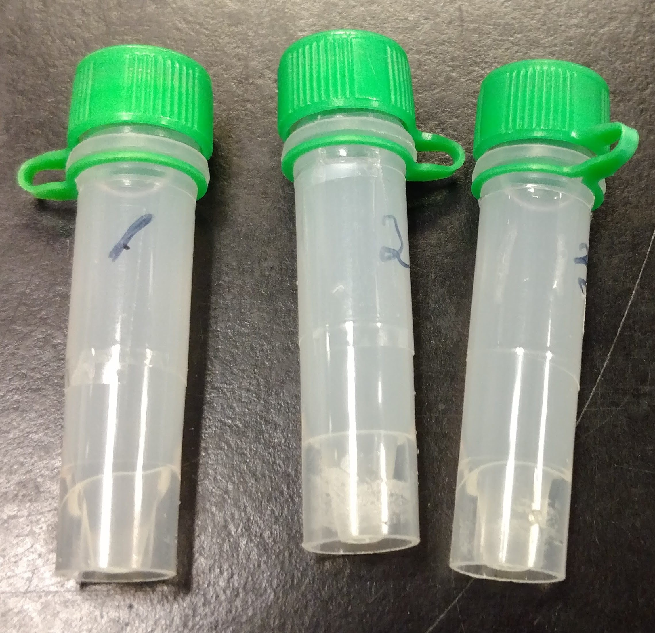
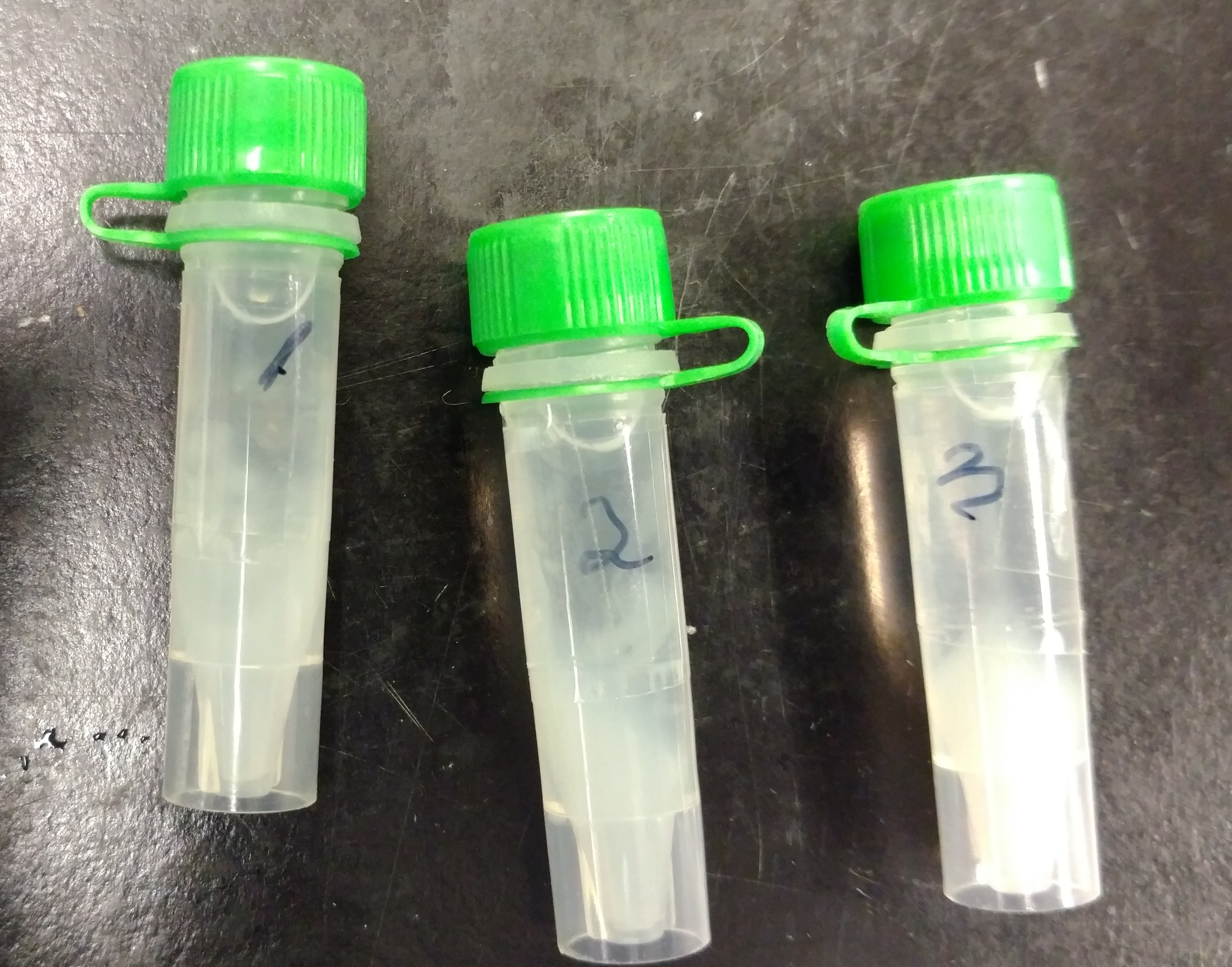
According to Jake’s notebook entry, the samples should have been previously homogenized in RNAzol RT. However, none of the samples showed evidence of being homogenized:

In theory, if these samples were snap frozen on liquid nitrogen after being placed in the RNAzol RT, there should be almost no impact on the RNA.
Procedure:
Samples were homogenized with disposable pestle in their respective tubes and vortexed.
Added 400μL of 0.1% DEPC-H2O to each sample and vortexed 15s.
Incubated samples 15mins at RT.
Centrifuged tubes 15mins at RT @ 16,000g.
750μL of the supe was transferred to a clean tube, added equal volume of isopropanol (750μL), mix by inversion (20 times), and incubated at RT for 15mins.
Centrifuged 12,000g for 10mins.
Discarded supe.
Washed pellets with 500μL of 75% EtOH (made with 0.1% DEPC-H2O) and centrifuged 4,000g for 3mins at RT. Repeated one time.
Removed EtOH and resuspended in 100μL of 0.1% DEPC-H2O. Most samples required vortexing to dissolve pellet.
Sample tubes were transferred to ice, quantified on the Roberts Lab NanoDrop1000, and stored @ -80C in their original box, pictured:

Results:


Google Spreadsheet with absorbance data: 20150506_Jake_Oly_1h_HS_RNA_ODs
Overall, the samples have excellent yields. The exceptions being the two samples that had less than 1mL of RNAzol RT in them to start (their yields are actually fine, but relative to all the other samples, they aren’t great). Should I have left them that way instead of adding additional RNAzol RT? Was there something wrong with these samples in the first place and that’s why they didn’t have a full 1mL of RNAzol RT in the tube already?
The 260/280 ratios are pretty good for most of the samples (>1.8), however I’d prefer to see RNA with 260/280 ratios >1.9.
The 260/230 ratios are amazing! The best I’ve seen coming straight out of an RNA isolation in a long time.
Eventually (once I’ve isolated RNA from the control set that corresponds to these heat shock samples), I’ll check for gDNA carryover and then, probably, DNase the RNA.