Tag Archives: triploid

Reverse Transcription – Ronit’s C.gigas DNased ctenidia RNA

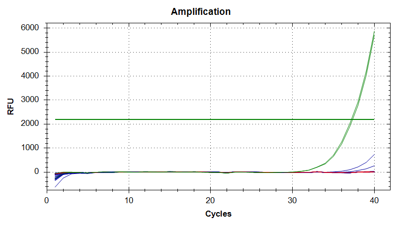
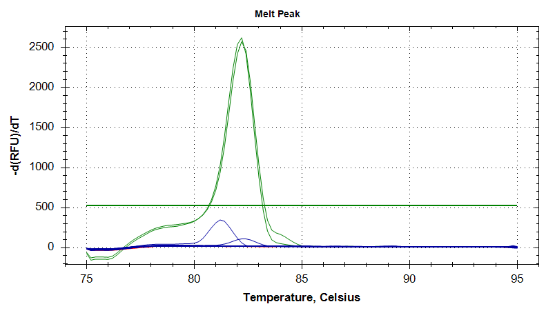
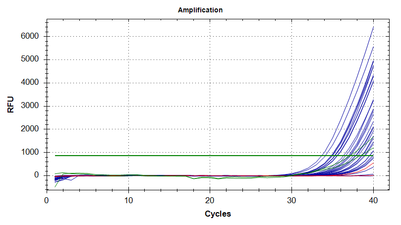
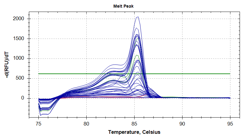
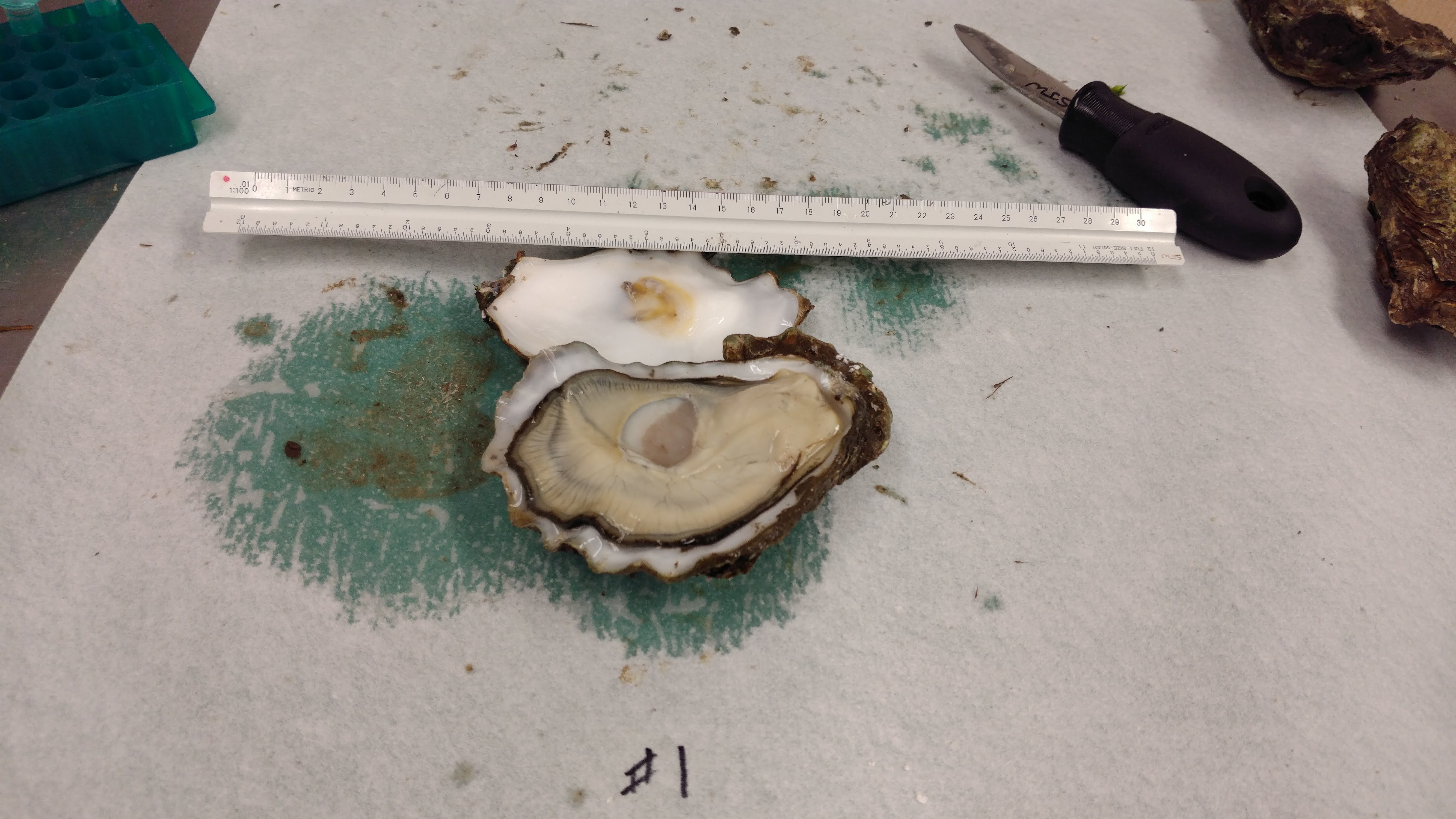
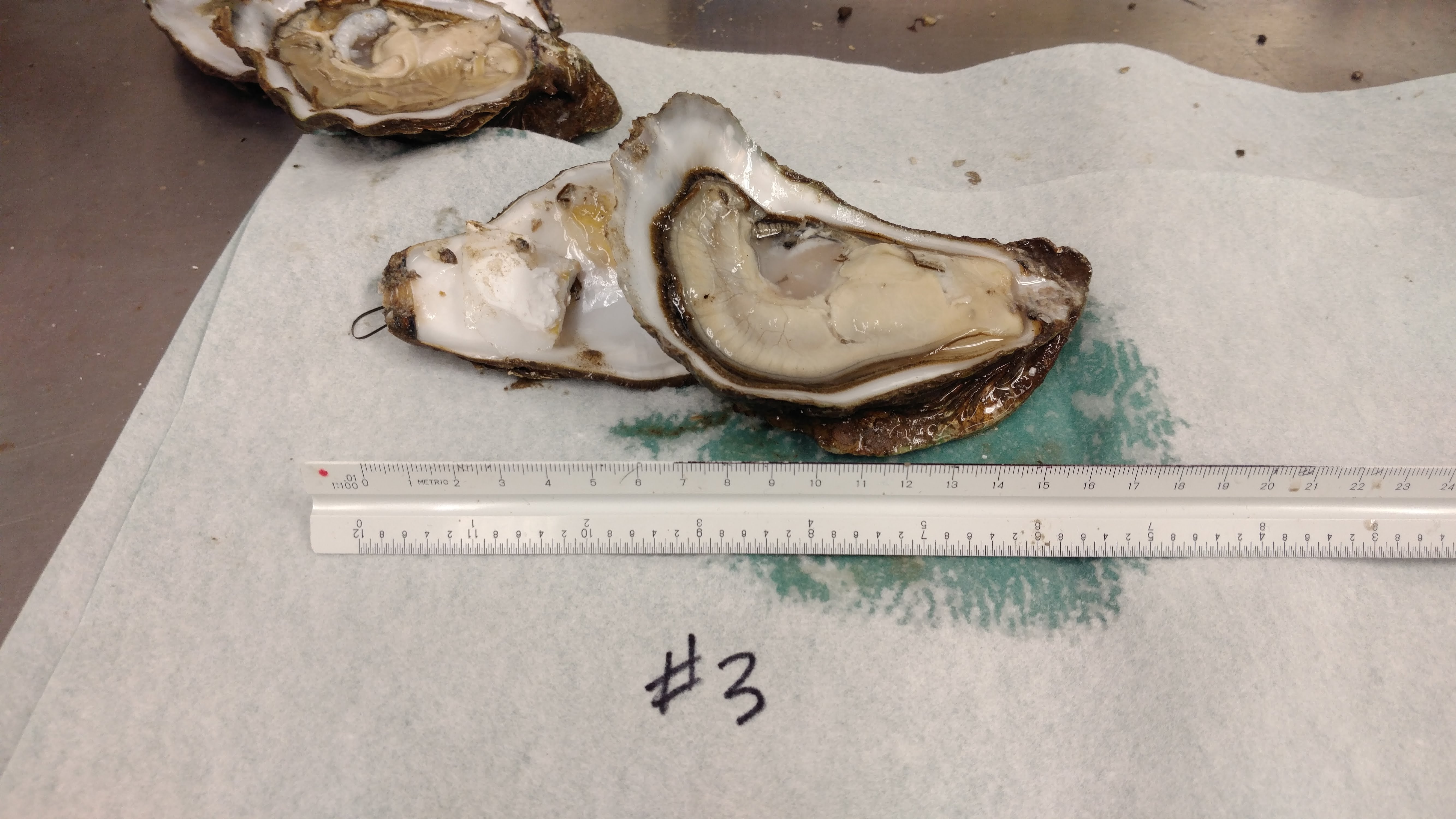
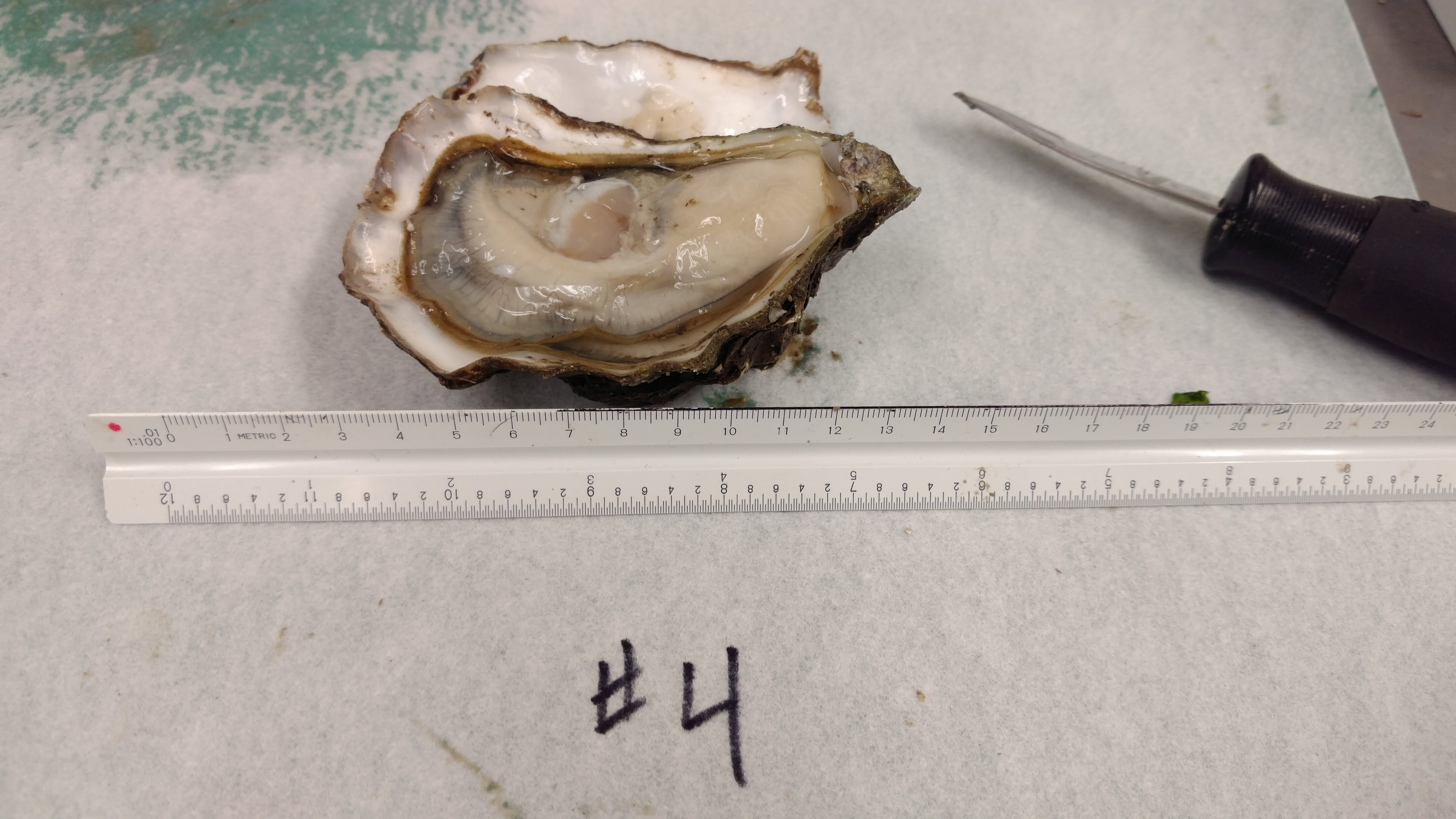
Proceeded with reverse transcription of Ronit’s DNased ctenidia RNA (from 20181016).

Reverse transcription was performed using 100ng of each sample with M-MMLV Reverse Transcriptase from Promega.

Briefly, 100ng of DNased RNA was combined with oligo dT primers and brought up to a final volume of 15uL. Tubes were incubated for 5mins at 70oC in a PTC-200 thermal cycler (MJ Research), using a heated lid. Samples were immediately placed on ice.

A master mix of buffer, dNTPs, water, and M-MMLV reverse transcriptase was made, 10uL of the master mix was added to each sample, and mixed via finger flicking. Samples were incubated for 1hr at 42oC in a PTC-200 thermal cycler (MJ Research), using a heated lid, followed by a 5min incubation at 65oC.

Samples were stored on ice for use later this afternoon by Ronit.

Samples will be stored in Ronit’s -20oC box.

Reverse transcription calcs (Google Sheet):

qPCR – Ronit’s DNAsed C.gigas Ploidy/Dessication RNA with elongation factor primers

After I figured out the appropriate DNA and primers to use to detect gDNA in Crassostrea gigas samples, I checked Ronit’s DNased ctenidia RNA (from 20181016) for residual gDNA.

Elongation factor primers:

  • EF1_qPCR_5′ (SRID 309)
  • EF1_qPCR_3′ (SRID 310)

BB16 from 20090519 was used as a positive control.

Samples were run on Roberts Lab CFX Connect (BioRad). All samples were run in duplicate. See qPCR Report (Results section) for plate layout, cycling params, etc.

qPCR master mix calcs (Google Sheet):


Results

qPCR Report (PDF):

qPCR File (PCRD):

qPCR Data (CSV):

In the plots below, green is the positive control, blue are the samples, and red is the no template control (NTC).

Everything looks great! Nice, clean, gDNA-free RNA! Will proceed with reverse transcription.


Amplification Plots


Melt Curves

qPCR – Ronit’s DNAsed C.gigas Ploidy/Dessication RNA with 18s primers

After DNasing Ronit’s RNA earlier today, I needed to check for any residual gDNA.

Identified some old, old C.gigas 18s primers that should amplify gDNA:

  • gigas18s_fw (SRID 157)
  • gigas18s_rv (SRID 156)

Used some old C.gigas gDNA (BB15 from 20090519) as a positive control.

Samples were run on Roberts Lab CFX Connect (BioRad). All samples were run in duplicate. See qPCR Report (Results section) for plate layout, cycling params, etc.

qPCR master mix calcs (Google Sheet):


Results

qPCR Report (PDF):

qPCR File (PCRD):

qPCR Data (CSV):

Well, this primer set and/or the gDNA is not good. In the plots below, the positive control gNDA is in green, samples in blue, and no template controls (NTC) are in red.

Poor performance is most easily noticed when looking at the melt curves. They have multiple peaks, suggesting non-specific amplification, even in the positive control.

Additionally, although less evident from just looking at the plots, is the replicates are highly inconsistent. Although it’s possible that might be due to poor technique, it’s very unlikely.

Will have to identify different primers and/or positive control DNA.


Amplification Plots


Melt Curves

DNase Treatment – Ronit’s C.gigas Ploiyd/Dessication Ctenidia RNA

After quantifying Ronit’s RNA earlier today, I DNased them using the Turbo DNA-free Kit (Ambion), according to the manufacturer’s standard protocol.

Used 1000ng of RNA in a 50uL reaction in a 0.5mL thin-walled snap cap tube. Samples were mixed by finger flicking and then incubated 30mins @ 37oC in a PTC-200 thermal cylcer (MJ Research), without a heated lid.

DNase inactivation was performed (0.1 volumes of inactivation reagent; 5uL), pelleted, and supe transferred to new 1.7mL snap cap tube.

Samples were stored on ice in preparation for qPCR to test for residual gDNA.

DNase calculations are here:

Samples will be permanently stored here (Google Sheet):

RNA Quantification – Ronit’s C.gigas Ploidy/Dessication RNA

Last Friday, Ronit quantified 1:10 dilutions of the RNA I isolated on 20181003 and the RNA he finished isolating on 20181011, but two of the samples (D11-C, T10-C) were still too concentrated.

I made 1:20 dilutions (1uL RNA in 19uL 0.1% DEPC-treated H2O) and quantified them using the Roberts Lab Qubit 3.0, with the RNA HS assay. Used 1uL of the diluted RNA.


RESULTS

Qubit data (Google Sheet):

Everything looks good. Added final concentration values (Qubit data x 20, to account for dilution factor) to Ronit’s master sheet (Google Sheet):

Will proceed with DNasing.

Samples Received – Triploid Crassostrea gigas from Nisbet Oyster Company

Received a bag of Pacific oysters from Nisbet Oyster Company.

Four oysters were shucked and the following tissues were collected from each:

  • ctenidia
  • gonad
  • mantle
  • muscle

Utensils were cleaned and sterilized in a 10% bleach solution between oysters.

Tissues were stored briefly on wet ice and then stored at -80C in Rack 2, Column 3, Row 1