Tag Archives: troubleshooting

qPCR – Test Young Lab qPCR Calibration

Recently, the Young Lab’s ABI 7300 qPCR machine was calibrated. Steven asked me to run a plate and see how well the calibration worked. Ran a plate with C.gigas gDNA and Gigas 18s primers (SR ID: 156 and 157) that are known to amplify gDNA. Master mix calcs are here (top half of page). Cycling params were as follows:

  • 95C – 10min

40 Cycles of:

  • 95C – 15s
  • 55C – 30s
  • 72C – 30s

Melt curve.

Results:

Absolutely no amplification of any kind. However, I did use one of our conventional PCR plates and not one of the ABI “prism” plates. Additionally, when I removed the plate from the machine, the plate looked as though it had been vigorously shaken:

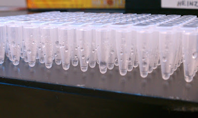
Will repeat this qPCR with a proper ABI “prism” plate.

qPCR – Test Plate for Opticon 2

Ran a full plate for testing well-to-well consistency (or, inconsistency!) of the Opticon 2, since it’s been behaving poorly lately. This will provide us with an idea of whether or not the oddities that we’ve been witnessing have any effect on our actual data.

Used C.gigas gDNA (DH15 from 20100519; 0.5128ug/uL) and IL17 Internal Fw/Rv primers (SR ID: 255, 256), which have previously produced an amplicon with gDNA. Master mix calcs/plate layout/cycling parameters/etc are here.

DNA was combined in master mix so that all wells received ~100ng of gDNA.

Results:

Opticon Calibration

Distributed 50uL of FAM calibration dye to wells. Ran out of dye!!

Looking back at old purchasing logs, it turns out we need 2 orders of dye packs to have enough for a 96-well plate.

Will cap existing plate with dye, wrap in foil and store @ -20C.

Ordered an additional pack of dye (Cat# 10006046; not available online, must call BioRad to order). Will ship on Monday. Will finish calibration procedure on Tuesday (20100928). Ugh.

Results:

Empty Plate:

Plate with dye (presumably calibrated):

After running the calibration protocol with the dye, all the wells should show consistent fluorescence levels. Clearly, they do not. Oddly enough, there appears to be a cyclical pattern across the wells of low -> high -> low fluorescence. The calibration protocol advises that if the wells do not exhibit consistent fluorescence across wells, then the plate should be read again. The graph above is the 2nd reading, which appears to be the same as the 1st. Conclusion is that the Opticon 2 is not working correctly and will contact BioRad for price quotes on repairs.

DNA Isolation – Qiagen Kit Comparison

Note: This information was added 20140407. Yes, you read that correctly.

Someone had noticed that gDNA isolated using a Qiagen DNeasy Blood & Tissue Kit we rec’d in April 2010 seemed to be yielding degraded DNA.

The two samples used for the comparison were a single tail (split in two equal weight pieces) from a juvenile salmon that was snap frozen, without preservatives, at the time of its collection. The samples were prepped. 0.5ug of eluted DNA was then run on a 1.2% agarose-TAE gel containing 0.1ug/mL of ethidium bromide. 5uL of Bioline’s Hyperladder I was loaded for size assessment (see link for marker layout).

Results:

Clearly, there’s significant quality difference! A free replacement kit was sent by Qiagen.

PCR – Test Lexie’s Mercenaria 18s contamination issue

Lexie’s PCR with this primer set and a pool of Mercenria cDNA has yielded contamination in all of her waters. Performed two sets of PCR: one with her existing primer working stocks and the other with a fresh aliquot of primer working stocks. Used my own reagents/water. PCR set up and cycling params are here. PCR ran O/N.

Results:

Lane 1 – 100bp ladder

Lane 2 – H2O (Lexie’s primer stocks)

Lane 3 – H2O (Lexie’s primer stocks)

Lane 4 – cDNA (Lexie’s primer stocks)

Lane 5 – H2O (fresh primer stocks)

Lane 6 – H2O (fresh primer stocks)

Lane 7 – cDNA (fresh primer stocks)

All water samples look clean and there’s a nice bright band in the cDNA samples. Lexie’s contamination issue is probably a technique issue and not one of reagent contamination.

qPCR – Gigas gDNA test of recalibrated Opticon 2

Master mix containing Gigas gDNA will be used to verify that the recalibration did work. qPCR setup/plate layout is here. I’ve made a master mix using Cg_HSP70_F/R primers designed by Mac. gDNA is BB #12 (0.445ug/uL) from 20090519. The gDNA will be added to the master mix.

Results: The results are a bit disconcerting, as this run shows virtually the exact same pattern in fluorescence detection as that on 20090722, despite using a different set of gigas gDNA. Below is a set of graphs comparing Column 1 Ct values of the two tests from 20090722 and today:

Clearly, both runs exhibit virtually the same pattern of relative Ct values to each other in each respective run. Not cool.

qPCR – Recalibration of Opticon 2

According to the Bio-Rad rep that analyzed the calibration test on 20090824, recalibration might be a good idea. As such, we’ll do that. Old calibration file was found and backed up prior to proceeding, as generation of a new calibration file would replace the old one.

Results: The Bio-Rad rep (Carl Fisher) has responded and said that the range of fluorescence is within the expected 2-fold difference and looks fine! Finally!

qPCR – Additional Calibration test of Opticon 2

Based on recs from Bio-Rad rep (Carl Fisher), will repeat Opticon 2 calibration (see 20090813) test according to Opticon manual. Then, will rotate plate 180 degrees and repeat test and upload data to Bio-Rad server for analysis and evaluation.

Results: According to the Bio-Rad rep, he thinks recalibration is in order. He pointed out that we’re seeing a 3-fold spread in fluorescence, which is outside of the “tolerable” range (which is 2-fold). Will recalibrate.

qPCR – Calibration test of Opticon 2

Received new FAM calibration reagent. It comes pre-prepared in a 1x PCR buffer (0.3uM), however there is only enough for a single plate (use 50uL/well). Will run plate in Opticon 2. Run according to the calibration protocol in the Opticon 2 manual (p. 10-4).

Results:

Well, there’s actually a signal this time as opposed to the run on 20090806. However, it’s pretty clear that the signals aren’t even close to being uniform. Or, as the manual says “tightly clustered lines”. I’m also not sure why the fluorescence decreases over time, although it could simply be degradation of the fluorophore after being hit with light. I’ve sent the results to Bio-Rad for help interpreting them.

qPCR – Calibration test of Opticon 2

Due to results of Opticon testing from 20090722, we have acquired FAM Calibartion Dye from Bio-Rad. Although not listed online or on the product itself, Bio-Rad customer service informed me that the concentration = 1mM. The calibration protocol in the Opticon 2 manual (p. 10-4) says to use 0.3uM (Cf) in 50uL. Will follow this info. Made up 15mLs of dye solution and distributed 50uL into each well of 3 plates. Tested all three plates on two different machines (Opticon 2 and Friedman lab’s).

Results: This did not produce any useable information whatsoever. In fact, the Friedman lab’s machine didn’t detect any signal at all from any of the three plates. Not sure what the story is.

UPDATE Received email correspondence from Carl Fisher of Bio-Rad and he has indicated that this is not the proper dye and that the conentration is not anywhere near 1mM (although he can’t find any info on what the concentration might be). He has told me to order a different part number (10006046) that is NOT listed anywhere on the Bio-Rad website.