Tag Archives: Qubit 3.0

DNA Isolation & Quantification – Metagenomics Water Filters

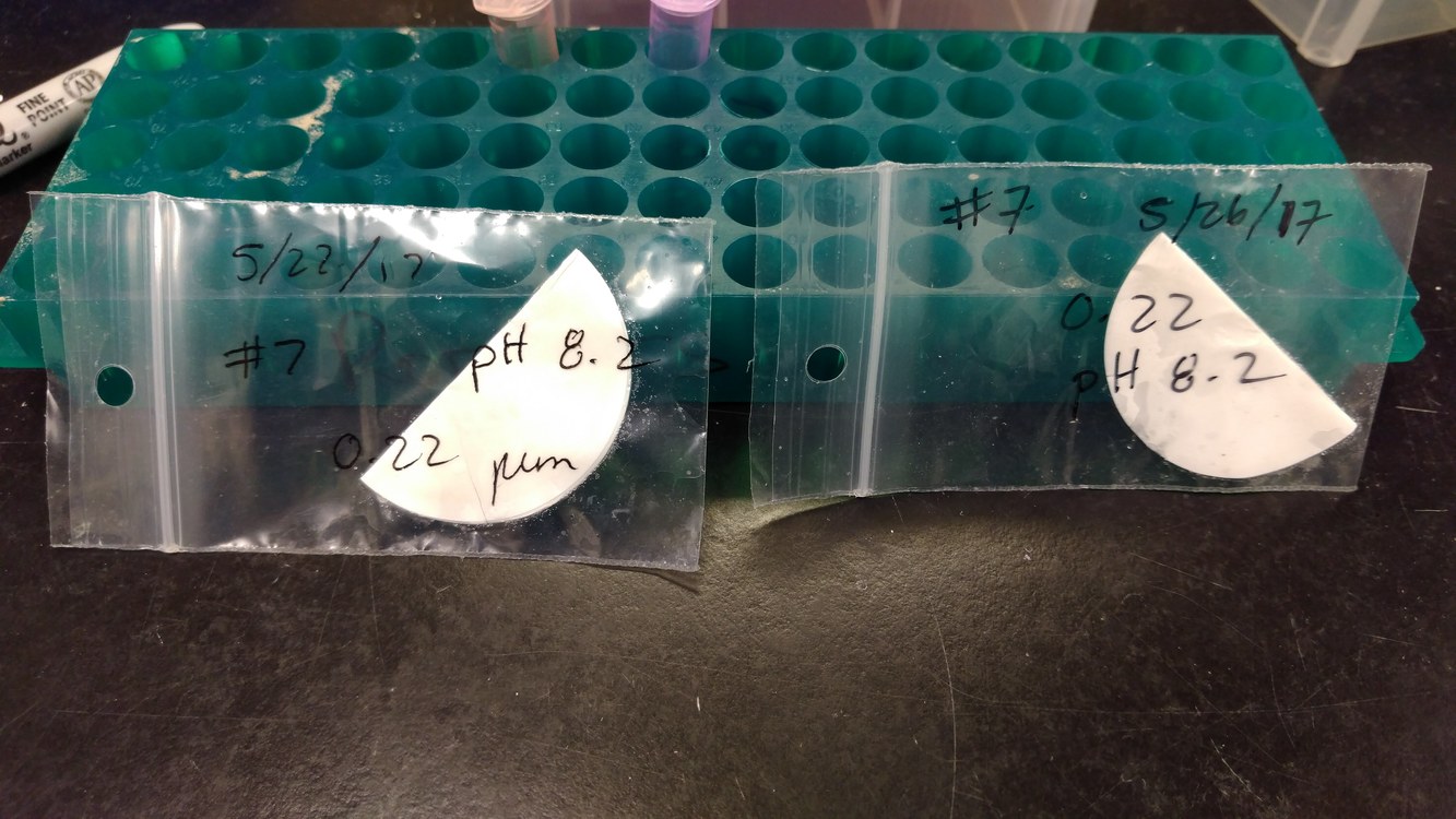
Isolated DNA from the following two filters:

DNA was isolated with the DNeasy Blood & Tissue Kit (Qiagen), following a modified version of the Gram-Positive Bacteria protocol:

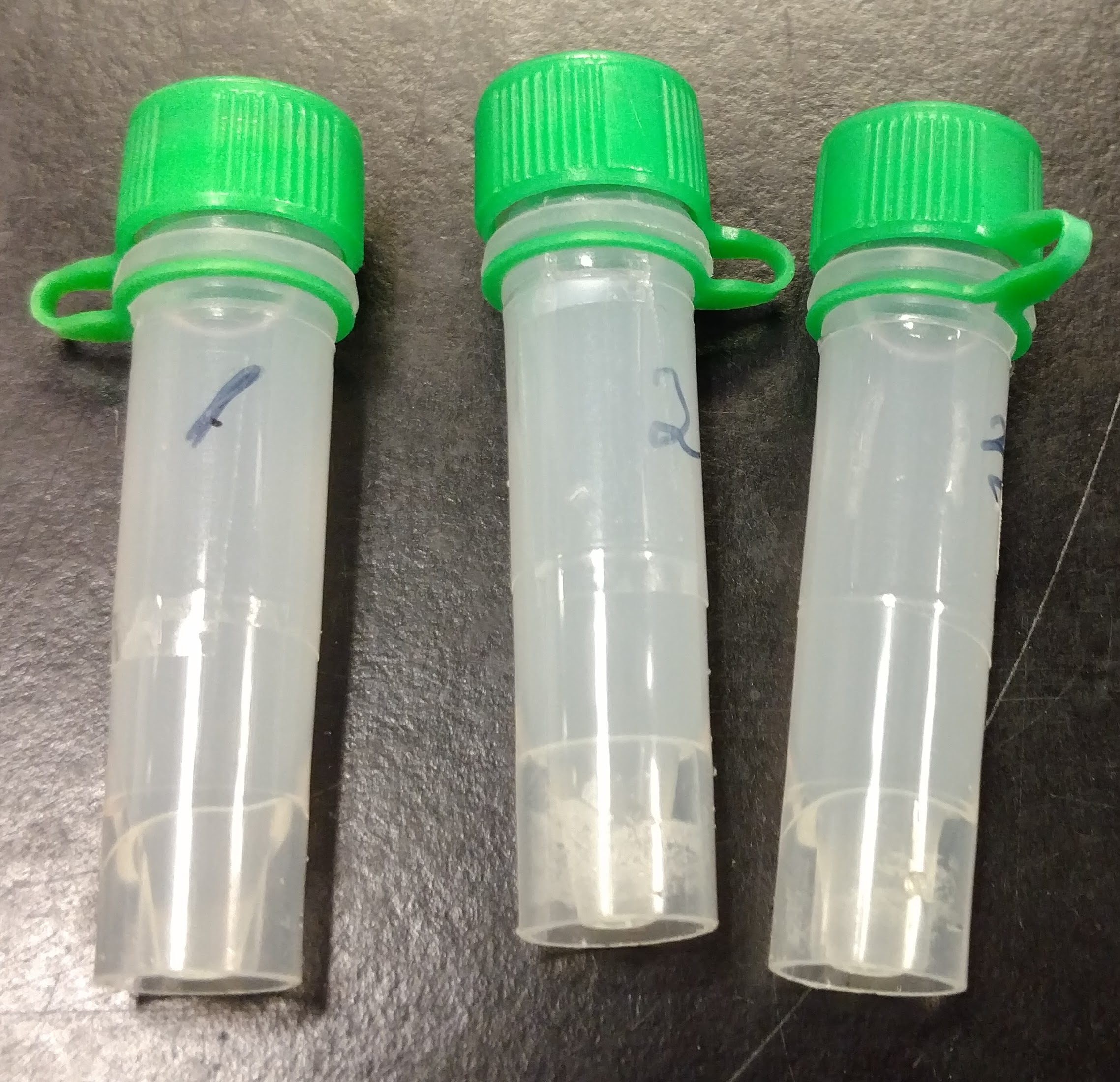
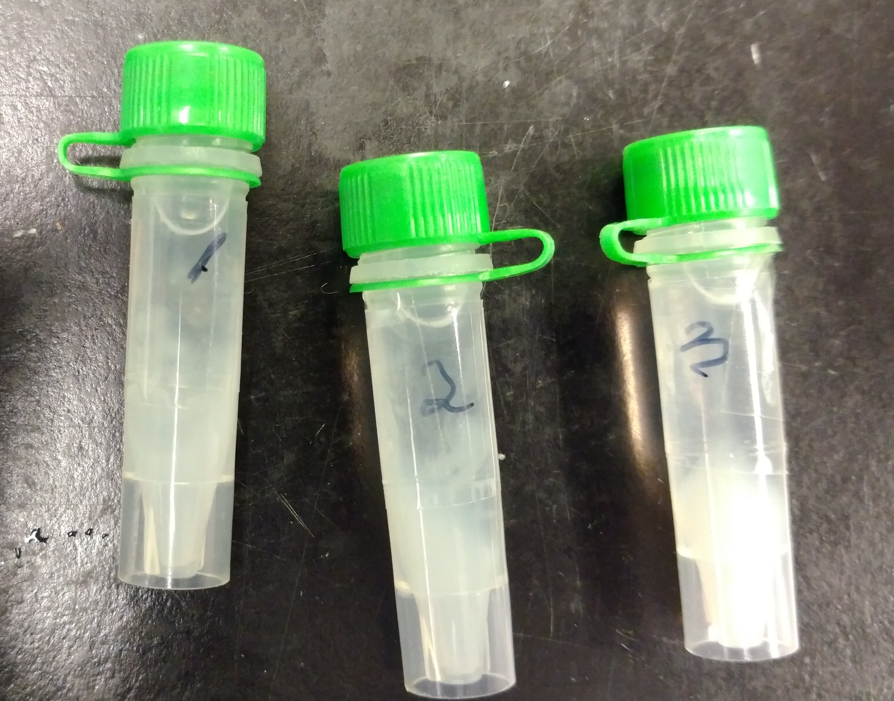
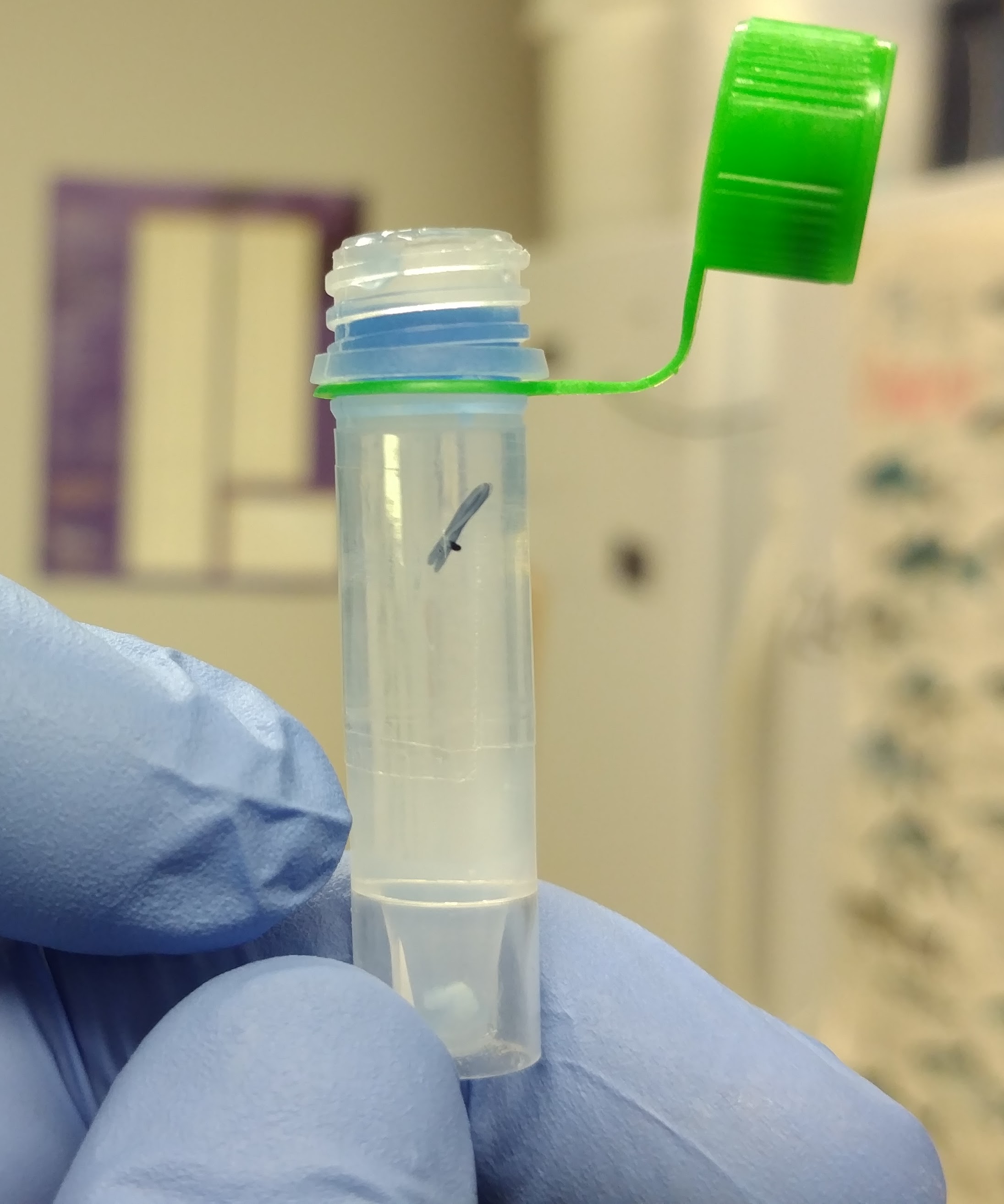
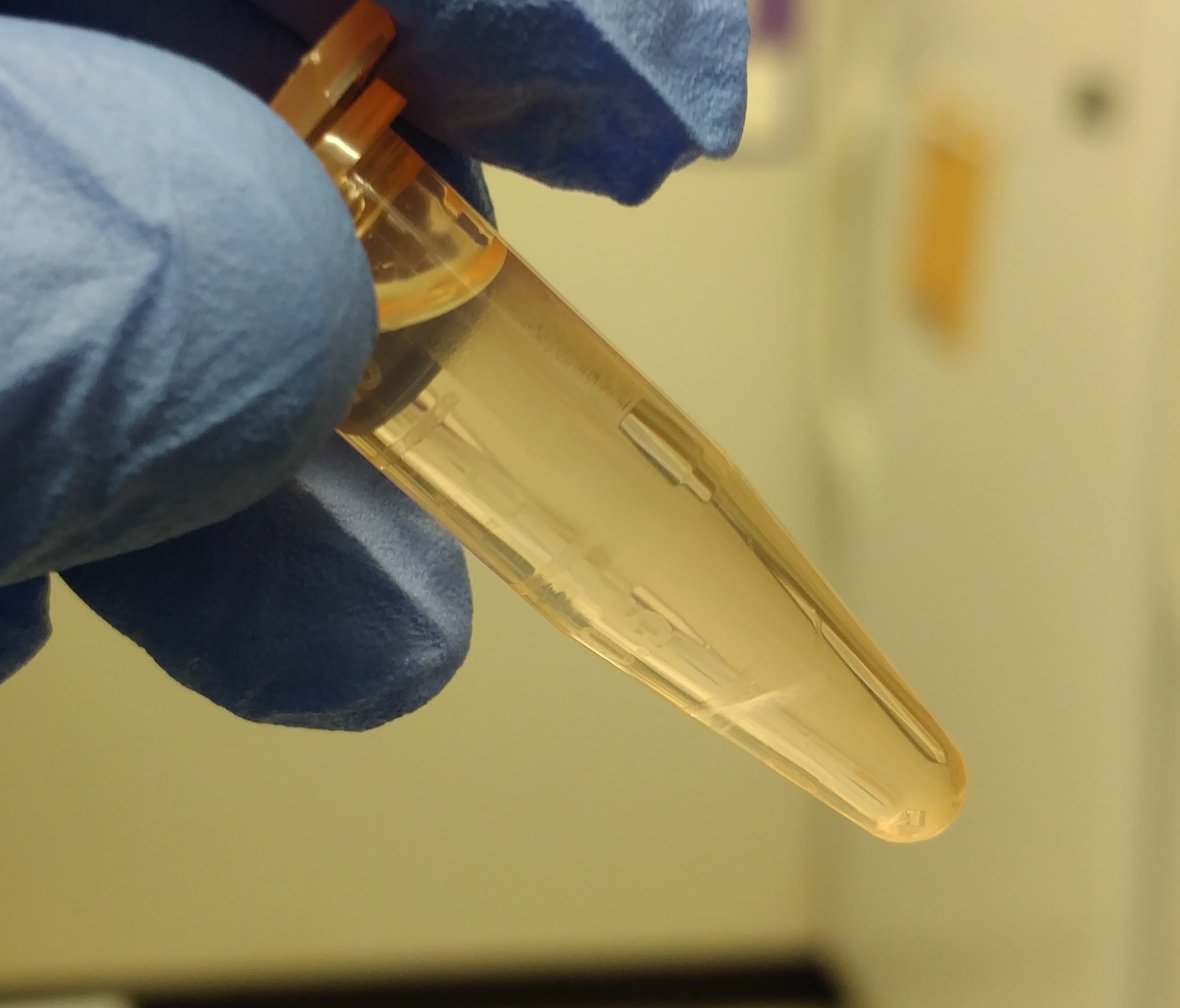
  • filters were unfolded and unceremoniously stuffed into 1.7mL snap cap tubes
  • did not perform enzymatic lysis step
  • filters were incubated with 400μL of Buffer AL and 50μL of Proteinase K (both are double the volumes listed in the kit and are necessary to fully coat the filter in a 1.7mL snap cap tube)
  • 56oC incubations were performed overnight
  • 400μL of 100% ethanol was added to each after the 56oC incubation
  • samples were eluted in 50μL of Buffer AE
  • all spins were performed at 20,000g

Samples were quantified with the Roberts Lab Qubit 3.0 and the Qubit 1x dsDNA HS Assay Kit.

Used 10μL of each sample for measurement (see Results for update).

Results:

Raw data (Google Sheet): 20180411_qubit_metagenomics_filters

Sample Concentration(ng/μL) Initial_volume(μL) Yield(ng)
filter 5/22 #7 pH8.2 20.8 50 1040
filter 5/26 #7 pH8.2 11.6 50 580

NOTE: For “filter 5/22 #7 pH8.2″ the initial quantification using 10μL ended up being too concentrated. Re-ran using 5μL.

Both samples have yielded DNA. This is, obviously, an improvement over the previous attempts to isolate DNA from ammonium bicarbonate filter rinses that Emma supplied me with.

Will discuss with Steven and get an idea of which filters to isolate additional DNA from.

Samples were stored Sam gDNA Box #2, positions G6 & G7. (FTR 213, #27 (small -20oC frezer)

DNA Isolation & Quantification – Geoduck larvae metagenome filter rinses

This is another attempt to isolate DNA from two more of the geoduck hatchery metagenome samples Emma delivered on 20180313.

The previous attempt, using DNAzol, did not yield any DNA.

I isolated DNA from the following two samples:

  • MG 5/19 #4
  • MG 5/26 #4

I used the DNA Stool Kit (Qiagen), following the “Stool Human DNA” protocol with the following changes:

  • Incubated @ 95oC for 5mins after initial addition of Buffer ASL. This is a lysis step that might help increase yields (see the “Stool Pathogen Detection” protocol)
  • Did not add InhibitEX Tablet. Deemed unnecessary, since these weren’t stool samples.
  • Eluted in 50μL of Buffer AE

I opted to follow the “Stool Human DNA” protocol, as it processes a larger portion of the initial sample, compared to the “Stool Pathogen Detection” protocol (600μL vs. 200μl)

Samples were quantified using the Roberts Lab Qubit 3.0 with the Qubit High Sensitivity dsDNA Kit (Invitrogen).

10μL of each sample were used.

Results:

Neither sample yielded any detectable DNA. Will discuss with Steven.

DNA Isolation & Quantification – Geoduck larvae metagenome filter rinses

Isolated DNA from two of the geoduck hatchery metagenome samples Emma delivered on 20180313 to get an idea of what type of yields we might get from these.

  • MG 5/15 #8
  • MG 5/19 #6

As mentioned in my notebook entry upon receipt of these samples, I’m a bit skeptical will get any sort of recovery, based on sample preservation.

Isolated DNA using DNAzol (MRC, Inc.) in the following manner:

  1. Added 1mL of DNAzol to each sample; mixed by pipetting.
  2. Added 0.5mL of 100% ethanol; mixed by inversion.
  3. Pelleted DNA 5,000g x 5mins @ RT.
  4. Discarded supernatants.
  5. Wash pellets (not visible) with 1mL 75% ethanol by dribbling down side of tubes.
  6. Pelleted DNA 5,000g x 5mins @ RT.
  7. Discarded supernatants and dried pellets for 5mins.
  8. Resuspended DNA in 20uL of Buffer EB (Qiagen).

Samples were quantified using the Roberts Lab Qubit 3.0 with the Qubit High Sensitivity dsDNA Kit (Invitrogen).

5uL of each sample were used.

Results:

As expected, both samples did not yield any detectable DNA.

Will discuss with Steven on what should be done with the remaining samples.

DNA Quantification – MspI-digested Crassostrea virginica gDNA

Quantified the two MspI-digested DNA samples for the Qiagen project from earlier today with the Qubit 3.0 (ThermoFisher).

Used the Qubit dsDNA Broad Range (BR) Kit (ThermoFisher).

Used 1μL of DNA from each sample (including undigested gDNA from initial isolation 20171211

Results:

Quantification (Google Sheet): 20180111_qubit_DNA_MspI_virginica

Yields are good and are sufficient for submission to Qiagen:

MspI_virginica_01 – 53.4ng/μL (1335ng; 89% recovery after phenol/chloroform/EtOH precip)
MspI_virginca_02 – 31.0ng/μL (775ng; ~52% recovery after phenol/chloroform/EtOH precip)

DNA Quantification – C.virginica MBD-enriched DNA

Quantified Crassostrea virginica MBD-enriched DNA from earlier today for Qiagen project.

Used the Qubit 3.0 (ThermoFisher) and the Qubit dsDNA Broad Range (BR) Kit (ThermoFisher).

Used 1uL of template DNA.

Results:

Quantification Spreadsheet (Google Sheet): 20180110_qubit_dsDNA_BR_MBD_virginica

Both samples had decent yields and have usable quantities for Qiagen (they wanted ~300ng from each sample):

virginica_MBD_01 – 18.3ng/uL (457.5ng = 5.7% methylated DNA capture)

virginica_MBD_02 – 19.6ng/uL (490ng = 6.1% methylated DNA capture)

Will store @ -20C until next week so that we’re not shipping so close to the weekend (shipping address is in Germany).

DNA Isolation & Quantification – Crassostrea virginica Mantle gDNA

DNA was isolated from a single adult Eastern oyster (Crassostrea virginica) for a pilot project with Qiagen to test their new DNA bisulfite conversion kit. The oyster was obtained yesterday afternoon (20171210) from the Taylo rShellfish Pioneer Square location. The oyster was stored @ 4C O/N.

The oyster was shucked and four pieces of upper mantle tissue (~35mg each) were snap frozen in liquid nitrogen (LN2). Tissues were pulverized under LN2 and then DNA was isolated separately from each sample using the E.Z.N.A. Mollusc DNA Kit (Omega) according to the manufcaturer’s protocol.

Samples were eluted with 100uL of Elution Buffer and were pooled into a single tube.

The gDNA was quantified using the Qubit 3.0 (Invitrogen) and Qubit dsDNA Broad Range Kit (Invitrogen), using 5uL of sample.

Results:

Qubit (Google Sheet): 20171211_qubit_virginica_DNA

Concentration is 58.4ng/uL.

That makes the total yield ~23.36ug (23360ng). This is more than enough to perform two separate MeDIP preps and two separate reduced representation digestions with MspI.

Will proceed with shearing of DNA for MeDIP.

DNA Isolation & Quantification – C. virginica Gonad gDNA

I isolated DNA from the Crassotrea virginica gonad samples sent by Katie Lotterhos using the E.Z.N.A. Mollusc Kit with the following modifications:

  • Samples were homogenized with plastic, disposable pestle in 350μL of ML1 Buffer
  • No optional steps were used
  • Eluted each in 100μL of Elution Buffer and pooled into a single sample

NOTE: Sample 034 did not process properly (no phase separation after 24:1 chlorform:IAA addition – along with suggested additions of ML1 Buffer) and was discarded.

Quantified the DNA using the Qubit dsDNA BR Kit (Invitrogen). Used 2μL of DNA sample.

Samples were stored in the same box the tissue was delivered in and stored in the same location in our -80C: rack 8, row 5, column 4.

Results:

Qubit (Google Sheet): 20171114_qubit_Cvirginica_gDNA

Ample DNA in all samples for MBDseq. (Refer to “Original Sample Conc.” column in spreadsheet.)

Will let Steven & Katie know.

RNA Isolation & Quantification – Tanner crab hemolymph

We received three Tanner crab (Chionoecetes bairdi)hemolymph samples from Pam Jensen (NOAA) yesterday. From her email to Steven:

Hi Steven,
I am sending:
tube #1 crab 3859/3656: 300 ul blood + 1300 ul RNAlater​

tube #2 crab 3665/3873: 300 ul blood + 1300 ul RNAlater
​tube #3 crab 3665/3873: 200 ul blood + 1400 ul RNAlater​

The tubes hold max of 1600 ul. Will know on Sun or Mon if either crab is infected w Hematodinium.

Tracking info to follow.
Pam

Samples were stored at 4C O/N.

Here’s what the samples looked like before processing:

The samples are extremely cloudy. I’m not sure if this is expected.

Processed samples using RNAzol RT (MRC) according to the manufacturer’s protocol for Total RNA Isolation.

Pelleted samples at 5000g for 5 mins and the samples looked like this:

Decided to pellet samples for an additional 10mins. The pellet was more compact. Transferred supernatant to clean tube, since it seemed to contain “debris” (maybe cells?). Processed pellet with RNAzol RT. Brief rundown of procedure (all steps at room temp):

  1. Transferred supe to clean tube.
  2. Added 1mL RNAzol RT to pellet and mixed by repeated pipetting (solution was cloudy and slightly viscous).
  3. Added 400uL of 0.1% DEPC-treated H2O and mixed vigorously by hand.
  4. Incubated for 10mins.
  5. Centrifuged 12,000g for 15mins.

    Samples looked like this:

    This is not normal. Usually the supernatant is the clear portion, while the blue layer is below that.
  6. Transferred 750uL of the clear portion to clean 1.7mL tube.

  7. Added equal volume of isopropanol, mixed by inversion. Appeared to be a very high amount of genomic DNA precipitation visible in the tube.
  8. Incubated for 10mins.
  9. Centrifuged 12,000g, 15mins.

    Samples looked like this:

    It appears that the nucleotides (the white interphase) are suspended on a “cushion” of higher density solution, instead of pelleted at the bottom of the tube.
  10. Removed/discarded higher density solution, leaving the white layer on the bottom of the tube.

  11. Centrifuged 12,000g, 15mins.
  12. Discarded supe.
  13. Washed pellet with 75% ethanol.
  14. Centrifuged 8,000g, 3mins.
  15. Repeated Steps 12, 13, & 14, 1x.
  16. Discarded ethanol.
  17. Resuspended RNA in 50uL 0.1% DEPC-treated H2O. Pellets did not solubilize on their own. I dispersed the pellets by repeated pipetting (P200). Remaining insoluble material was pelleted (12,000g, 30s) and supernatant was transferred to a new 1.6mL tube.

RNA was quantified using the Qubit 3.0 and the Qubit HS RNA Assay. Used 5uL of each sample.

Results:

20171107_qubit_tanner_crab_hemo (Google Sheet)

Sample ID Conc. (ng/uL) Total Yield (ng)
3859/3656 0.44 22
3665/3873 1.66 83
3665/3873 2.04 102

Interestingly, both samples from the same crab had similar/decent yields.

Samples were labeled and stored at -80C in Shellfish RNA Box #6

RNA Isolation – Olympia oyster gonad tissue in paraffin histology blocks

UPDATE 20170712: The RNA I isolated below is from incorrect regions of tissue. I misunderstood exactly what this tissue was, and admittedly, jumped the gun. The tissue is actually collected from the visceral mass – which contains gonad (a small amount) and digestive gland (a large amount). The RNA isolated below will be stored in one of the Shellfish RNA boxes and I will isolate RNA from the correct regions indicated by Grace

Isolated RNA from Olympia oyster gonad previously preserved with the PAXgene Tissue Fixative and Stabilizer and then embedded in paraffin blocks. See Laura’s notebook for full details on samples and preservation.


RNA was isolated from the following samples using the PAXgene Tissue RNA Kit (Qiagen). Gouged samples from the blocks weighing ~10mg from each of the tissues and processed according the protocol for isolating RNA from blocks of paraffin-embedded tissues.

Tissue identification is available in this GitHub Issue

NF-10-22
NF-10-23
NF-10-24
NF-10-26
NF-10-28
NF-10-30
SN-10-16
SN-10-17
SN-10-20
SN-10-25
SN-10-26
SN-10-31

IMPORTANT:

  • Prior to beginning, I prepared an aliquot of Buffer TR1 by adding 40μL of β-mercaptoethanol (β-ME) to 4000μL of Buffer TR1).
  • Reconstituted DNase I with 550μL of RNase-free H2O. Aliquoted in 100μL volumes and stored @ -20C in the “-20C Kit Components” box.

Isolated RNA according to the PAXgene Tissue RNA Kit protocol with the following alterations:

  • “Max speed” spins were performed at 20,000g.
  • Tissue disruption was performed by adding ~25-50 glass beads (425 – 600μm diameter) with the Disruptor Genie @ 45C for 15mins (in the Friedman Lab).
  • Shaking incubation step was performed with Disruptor Genie
  • Samples were eluted with 27μL of Buffer TR4 x 2, incubated @ 65C for 5mins, immediately placed on ice and quantified on the Roberts Lab Qubit 3.0 with the RNA High Sensitivity Assay (ThermoFisher Scientific) using 5μL of each sample.

Results:

Concentrations (Google Sheet): 20170710_RNA_qubit_oly_histo_blocks

Well, the good news is that there’s RNA from all the samples and it seems to be in relatively high concentrations!

The bad news is that the concentrations for 10 of the 12 samples were too high and outside the range of the Qubit RNA HS Assay! Since we don’t have the broad range RNA assay, I can’t properly quantify the remaining samples. However, these samples are being sent to Katherine Silliman at some point, so I’ll leave it up to her to quantify the samples. I’m also guessing that she’ll run them on a Bioanalyzer to assess their integrity prior to beginning library construction, so that will also yield concentrations for the samples.

Samples were stored at -80C temporarily.

Samples will be sent to Katherine Silliman for high-throughput library construction and sequencing once I hear back from her regarding her availability to receive the samples.