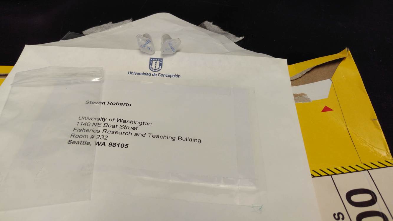
Received Caligus tape DNA – two samples:
- Female 1 .
-
Female 2 .
Stored in slots H4 and H5 in “Sam’s gDNA Box #2″ in the FTR 213 -20oC freezer.
Google Sheet: Sam’s gDNA Box #2
Received Caligus tape DNA – two samples:
Female 2 .
Stored in slots H4 and H5 in “Sam’s gDNA Box #2″ in the FTR 213 -20oC freezer.
Google Sheet: Sam’s gDNA Box #2
Used the MethylFlash Methylated DNA Quantification Kit (Colorimetric) from Epigentek to quantify methylation in these coral DNA samples.
All samples were run in duplicate <em>except</em> 2h Block 1 due to insufficient DNA.
The following samples were used in a 1:10 dilution (2uL DNA : 18uL NanoPure H2O), due to their relatively high concentrations, to ensure accurate pipetting:
All samples were diluted to a final concentration of 9.645ng/uL (154.24ng total; 17.6uL) in NanoPure water, which is equal to 77.12ng of DNA per assay replicate. These numbers were chosen based off of the sample with the lowest concentration.
The following samples were used in their entirety:
Calculations were added to the spreadsheet provided by Javier (Google Sheet): A.cervicornis_DNA_Extractions(May_2017).xlsx
The spreadsheet became overly complicated because I initially forgot to account for the need to run each sample in duplicate.
The kit reagent dilutions were as follows:
All diluted solutions were stored on ice for duration of procedure.
The remaining Diluted ME1 solution was stored at 4C (FTR 209), and is stable for 6 months, per the manufacturer’s instructions.
See the Results section below for plate layouts.
Plates were read at 450nm on the Seeb Lab Victor 1420 Plate Reader (Perkin Elmer) and the amount of DNA methylation was determined.
Results:
Individual sample methylation quantification (Google Sheet): A.cervicornis_DNA_Extractions(May_2017).xlsx
Plate Reader Output File Plate #1 (Google Sheet): 20170511_coral_DNA_methylation_plate01.xls
Plate Reader Output File Plate #2 (Google Sheet): 20170511_coral_DNA_methylation_plate02.xls
I’m not familiar with the experimental design, so I’m not going to spend time handling any of the in-depth analysis at this point in time. However, here’s the background on how methylation quantification and percent methylation were determined.
Mean absorbance (450nm) was determined for all samples and standard curve samples. It’s important to note that the standard deviation between replicates was not evaluated and there appears to be consistent variability between samples, but I’m not certain how much variation is “acceptable” with and assay of this nature.
The mean absorbance of the standard curve samples were plotted against their corresponding DNA amounts and a linear trendline was fitted to the points.
Per the manufacturer’s recommendations, the four points (including the zero point) that yielded the best linear fit (i.e. best R^2 value) were used and the slope of best fit line for those four points was determined.
This slope was then utilized in the equation provided by the manufacturer (see pg. 8 of the MethylFlash Kit manual).
Received 62 coral (Acropora cervicornis) DNA samples from Javier Casariego at FIU.
Spreadsheet of samples and NanoDrop concentrations provided by Javier (converted to Google Sheet): A.cervicornis_DNA_Extractions(May_2017).xlsx
Samples were temporarily stored at 4c (in FTR 213) until I can perform global methylation assessment on them tomorrow.

Ran the coral DNA I quantified on 20160630 through the MethylFlash Methylated DNA Quantification Kit [Colorimetric] (Epigentek) kit to quantify global methylation.
Used 100ng of DNA per 8uL per replicate (x2 replicates = total 200ng in 16uL). Calcs are here (Google Sheet): 20160705_coral_DNA_methylation_calcs
Manufacturer’s protocol was followed.
Dilutions of kit reagents:
ME5 (1:1000) 2.6uL ME5 + 2597.4uL diluted ME1
ME6 (1:2000) 1.3uL ME6 + 2598.7uL diluted ME1
ME7 (1:5000) 0.52uL ME7 + 2599.48uL diluted ME1
Samples were quantified on the Seeb’s plate reader @ 450nm (Wallac 1420 Victor 2 [Perkin Elmer])
Results:
Google Sheet: 20160707_coral_DNA_methylflash
| sample | treatment | 5-mC(ng) |
| H1_1 | nitrogen | 0.8712248853 |
| H1_10 | nitrogen | 0.6917168368 |
| H1_12 | control | 0.2738478893 |
| H1_5 | nitrogen & phosphorous | 0.9663585942 |
| H1_6 | control | 0.6494783825 |
| H1_8 | nitrogen & phosphorous | 0.4244913398 |
| H24_1 | nitrogen | 0.372603297 |
| H24_10 | nitrogen | 0.4237237786 |
| H24_12 | control | 0.5350511937 |
| H24_5 | nitrogen & phosphorous | 0.1495527697 |
| H24_6 | control | 0.2291900804 |
| H24_8 | nitrogen & phosphorous | 0.2213437801 |
| H5_1 | nitrogen | -0.1233169902 |
| H5_10 | nitrogen | 0.6997668774 |
| H5_12 | control | 0.2307000493 |
| H5_5 | nitrogen & phosphorous | -0.07790933048 |
| H5_6 | control | 0.4562401662 |
| H5_8 | nitrogen & phosphorous | 0.5949647121 |
Overall, it’s difficult to really interpret these results. I believe the data is a time course (e.g. H5 = hour 5, H24 = hour 24). Additionally, looking at treatments, there appear to be replicates, but it’s not clear what type of replicates they are (i.e. technical or biological). Generally, it seems like the control samples have lower quantities of methylated DNA than the treated samples. However, this doesn’t hold true for all three of the groups.
And, not that it really matters, but I don’t even know what species this is…
In any case, this was an attempt to gather some preliminary data for a grant that Steven is attempting to put together, so the original experiment and the subsequent data aren’t as robust as one would expect for a full-blown research project.
Quantified the DNA we received from Jose on 20160615 using the Qubit 3.0 Flouorometer (ThermoFisher) with the dsDNA Broad Range (BR) Kit according to the manufacturer’s protocol. Used 1μL of each sample.
Results are here (Google Sheet): Coral_DNA_QubitData_2016-06-30_08-45-56.xls
Here is a table of sample concentrations:
| Sample | Concentration(ng/μL) |
| H1 1 | 52.4 |
| H1 5 | 34 |
| H1 6 | 13 |
| H1 8 | 22 |
| H1 10 | 39 |
| H1 12 | 52.4 |
| H5 1 | 14.7 |
| H5 5 | 20.8 |
| H5 6 | 54 |
| H5 8 | 18.4 |
| H5 10 | 46.6 |
| H5 12 | 29.8 |
| H24 1 | 16.2 |
| H24 5 | 25 |
| H24 6 | 20.2 |
| H24 8 | 22 |
| H24 10 | 22 |
| H24 12 | 30.6 |
Will proceed with DNA methylation assessment.
Steven received these coral DNA samples today. Here’s his post on Google Plus (stored @ 4C in FTR 213):
Here’s the email from Jose describing the samples:
“Dear Steven, the coral DNA samples were sent today by my student Javier (cc’ed here) to your lab. Here’s an excel attached with info for the samples including concentration and treatment of the coral from which they were extracted (N, nitrogen; NP, nitrogen+phosphorous; C, control).
Please let us know when you get these in the lab so we know all is fine!
thanks!
Chema”
Here’s the spreadsheet he sent (renamed for easier identification later on – original file sent was title DNA Qbit readings), uploaded to Google Drive:
Purified DNA from the remaining PCR bands excised by Jake on 20150609 and 20150610, as well as Jonathan’s sea pen PCRs from 20150604, using Ultrafree-DA spin columns (Millipore). Transferred gel pieces from storage tubes (1.5mL snap cap tubes) to spin columns. Spun 10,000g, 5mins @ RT. Transferred purified DNA back to original storage tubes. See the sequence_log (Google Sheet) for a full list of the samples and the sequencing plates layouts. Purified DNA was stored @ 4C O/N. Will prepare and submit plates for Sanger sequencing tomorrow.
After the discovery that there wasn’t any DNA in the BS-seq Illumina library prep and no DNA in the bisulfite-treated DNA pool, I decided to try to recover any residual DNA left in the 1B2 sample. Sample 1B2 (sheared on 20150109) was dry, so I added 20μL of Buffer EB (Qiagen) to the tube. I vortexed both the 1B1 and 1B2 samples and quantified on the NanoDrop1000 (ThermoFisher). I also re-quantified the pooled BS-treated sample that had been used as input DNA for the libraries.
Results:
Spreadsheet: 20150226_Claire_sheared_Emma_1000ppm_OD260s
Sample 1B1 has ample DNA in it. Since these samples are pools of larvae, we may be able to just proceed with this sample and not worry about pooling with the biological replicate 1B2.
Sample 1B2 has a low amount of DNA, but it’s a usable quantity (total 400ng).
Pooled samples has nothing.
Will make a new pool of DNA from both 1B1 and 1B2 and attempt to make a new bisulfite-treated library.
To complement MBD ChiP-seq data and RNA-seq data that we have from this experiment, we want to generate, at a minimum, some BS-seq data from the same C.gigas individuals used for the other aspects of this experiment. Claire had previously isolated DNA and sheared the DNA on 20140108. If possible, we’d like to perform MBD enrichment, but the current quantities of DNA may prevent us from this.
To quantify the DNA and evaluate the shearing profile, I ran 1μL of each of the following mantle pre-/post-heat shock samples on a DNA 1000 chip (Agilent) on the Agilent 2100 Bioanalyzer. in the Seeb Lab:
M = mantle
HS = heat shocked
Results:
Bioanalyzer Data File (XAD): 2100_expert_DNA_1000_DE72902486_2015-02-19_11-32-35(2).xad
Electropherograms

2100 Bioanalyzer electropherograms of Claire’s sheared C.gigas DNA.
Spreadsheet: 2100 expert_DNA 1000_DE72902486_2015-02-19_11-32-35_Results_001
Claire’s notebook entry doesn’t ever specify what her target shear size was, but the Bioanalyzer analysis suggests an average size of ~500bp.
Also interesting to note is that Claire’s sample concentrations (as measured on the NanoDrop1000) are significantly greater than what is calculated by the Bioanalyzer. Since the Bioanalyzer chip used (DNA1000) only goes to 1000bp, is it possible the differences in concentrations is due to incomplete shearing of the samples (e.g. a significant portion of the DNA is >1000bp in size and thus not factored in to the Bioanlyzer concentrations calculations)?
Will check sample volumes and determine total amount of remaining DNA for each sample and then assess how to proceed next (i.e. MBD or just BS-seq).
UPDATE 20150226:
Sample volumes were measured and total quantity (ng) of DNA in each sample were added to the spreadsheet above.
Based on the quantities of DNA we have for each sample, will discuss sequencing options (e.g. MBD or not, self-prepare libraries or not, etc) with Steven.

